. 2012 May;22(5):1016-24.
doi: 10.1093/cercor/bhr176. Epub 2011 Jul 19.

The effect of preterm birth on thalamic and cortical development

Affiliations

The effect of preterm birth on thalamic and cortical development

Gareth Ball et al. Cereb Cortex. 2012 May.

Abstract

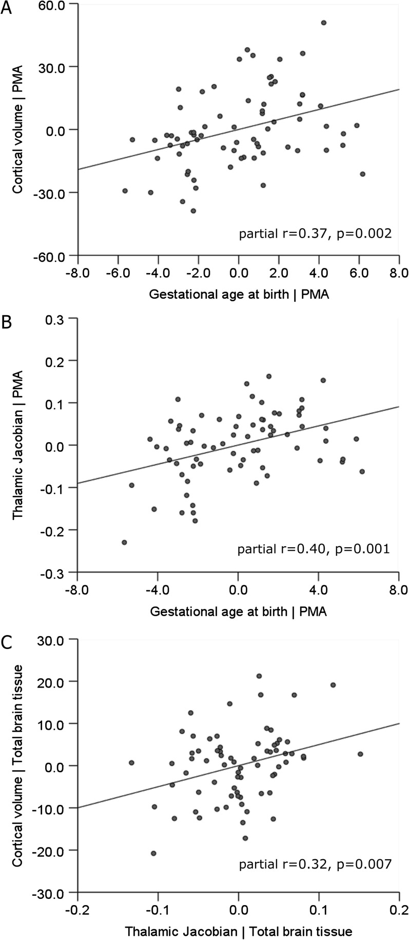
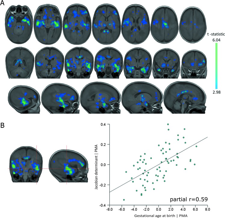
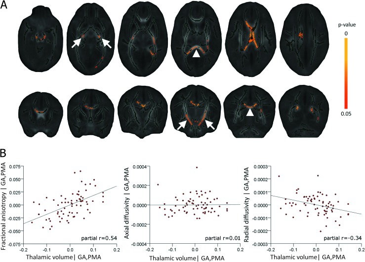
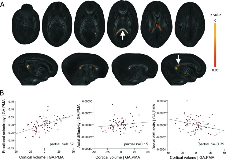
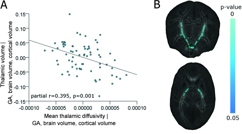
Preterm birth is a leading cause of cognitive impairment in childhood and is associated with cerebral gray and white matter abnormalities. Using multimodal image analysis, we tested the hypothesis that altered thalamic development is an important component of preterm brain injury and is associated with other macro- and microstructural alterations. T(1)- and T(2)-weighted magnetic resonance images and 15-direction diffusion tensor images were acquired from 71 preterm infants at term-equivalent age. Deformation-based morphometry, Tract-Based Spatial Statistics, and tissue segmentation were combined for a nonsubjective whole-brain survey of the effect of prematurity on regional tissue volume and microstructure. Increasing prematurity was related to volume reduction in the thalamus, hippocampus, orbitofrontal lobe, posterior cingulate cortex, and centrum semiovale. After controlling for prematurity, reduced thalamic volume predicted: lower cortical volume; decreased volume in frontal and temporal lobes, including hippocampus, and to a lesser extent, parietal and occipital lobes; and reduced fractional anisotropy in the corticospinal tracts and corpus callosum. In the thalamus, reduced volume was associated with increased diffusivity. This demonstrates a significant effect of prematurity on thalamic development that is related to abnormalities in allied brain structures. This suggests that preterm delivery disrupts specific aspects of cerebral development, such as the thalamocortical system.


Figures

Figure 1.
Figure 1.
Final reference template and thalamic and cortical segmentations. The final average intensity template is shown in (A), the clarity of the subcortical structures and cortical differentiation indicates the accurate alignment of individual images. The mean Jacobian determinant within the mask shown in (B) represents the relative volume change between the template and each image and was used to represent thalamic volume across the cohort. A representative example of cortical gray matter segmentation is shown in (C).
Figure 2.
Figure 2.
DBM processing pipeline. After preprocessing, T1 images are affinely aligned to an arbitrarily chosen target MR image and averaged to produce a reference template. Two subsequent iterations of nonlinear registration and template construction produce the final transformations used for analysis.
Figure 3.
Figure 3.
Cortical gray matter volume is correlated with prematurity at birth and thalamic volume at term-equivalent age. Partial regression plots show significant associations between cortical gray matter volume and gestational age at birth (A) and mean thalamic Jacobian (representing thalamic volume) and gestational age (B), after correction of each measure for the postmenstrual age at scan (PMA) of each infant. Shown in (C) is the significant association between cortical volume and thalamic Jacobian, after correction of each for total cerebral tissue volume.
Figure 4.
Figure 4.
Regional associations between brain tissue volume and prematurity at birth. Regions where tissue volume is significantly associated with gestational age at birth after correcting for the age of each infant at scan are shown in (A). Statistical images are corrected for multiple comparisons at P < 0.01 FDR-corrected (color bar indicates t-statistic). To illustrate this relationship, the Jacobian determinant, representing volume change relative to the reference template, at the site of the maximum t-statistic (red crosshairs; t = 6.04), was entered into a multiple linear regression with gestational age at birth and age at scan. The partial regression plot (B) shows the relationship between Jacobian and gestational age at birth.
Figure 5.
Figure 5.
Regional associations between brain tissue volume and thalamic volume at term-equivalent age. Regions where cerebral tissue volume significantly covaried with mean thalamic Jacobian (calculated within the region circled in red). Arrows indicate the hippocampi, statistical images are corrected for multiple comparisons at P < 0.001 FDR-corrected (color bar indicates t-statistic).
Figure 6.
Figure 6.
Thalamic volume is associated with white matter microstructure. Regions where fractional anisotropy is significantly associated with thalamic volume, beyond any common association with prematurity at birth and age at imaging, are shown in (A). These regions include the posterior limb of the internal capsule (arrows) and the corpus callosum (arrowheads). Images are FWE-corrected at P < 0.05 (color bar indicates P value), the mean FA skeleton is shown in dark green. Partial regression plots of the relationship between thalamic volume and mean FA, axial diffusivity (AD), and radial diffusivity (RD) extracted from each significant voxel identified in (A) and entered into linear regression with gestational age and age at scan are shown in (B).
Figure 7.
Figure 7.
Cortical volume is associated with white matter microstructure. Fractional anisotropy in the posterior corpus callosum (A; arrow, bottom row) including the splenium (A; arrow, top row) is significantly associated with cortical volume, after correction for prematurity at birth and age at imaging. Images are shown as in Figure 6. Cortical volume and mean FA, AD, and RD extracted from each significant voxel identified in (A) were entered into linear regression with gestational age at birth (GA) and age at scan (PMA). Partial regression plots of the relationship between cortical volume and FA, AD and RD, after correction for gestational age and age at scan are shown in (B).
Figure 8.
Figure 8.
Thalamic diffusivity is associated with thalamic volume and FA in the internal capsule. Thalamic volume (estimated from the mean Jacobian) and mean thalamic diffusivity (estimated from a thalamic mask placed in the DTI reference space) were entered into a multiple linear regression model with gestational age at birth (GA), cortical volume, and total brain volume. The partial regression plot in (A) shows the significant association between thalamic volume and thalamic diffusivity. In (B), regions where FA was significantly associated with thalamic diffusivity are shown (FWE-corrected at P < 0.05), beyond any associations with GA, PMA, cortical volume, and CLD status.

Similar articles

Cited by

References

    1. Ajayi-Obe M, Saeed N, Cowan FM, Rutherford MA, Edwards AD. Reduced development of cerebral cortex in extremely preterm infants. Lancet. 2000;356:1162–1163. - PubMed
    1. Allendoerfer KL, Shatz CJ. The subplate, a transient neocortical structure: its role in the development of connections between thalamus and cortex. Annu Rev Neurosci. 1994;17:185–218. - PubMed
    1. Anjari M, Counsell SJ, Srinivasan L, Allsop JM, Hajnal JV, Rutherford MA, Edwards AD. The association of lung disease with cerebral white matter abnormalities in preterm infants. Pediatrics. 2009;124:268–276. - PubMed
    1. Anjari M, Srinivasan L, Allsop JM, Hajnal JV, Rutherford MA, Edwards AD, Counsell SJ. Diffusion tensor imaging with tract-based spatial statistics reveals local white matter abnormalities in preterm infants. Neuroimage. 2007;35:1021–1027. - PubMed
    1. Arai Y, Deguchi K, Mizuguchi M, Takashima S. Expression of beta-amyloid precursor protein in axons of periventricular leukomalacia brains. Pediatr Neurol. 1995;13:161–163. - PubMed

Publication types