Review
. 2011 Aug 1;187(3):1081-9.
doi: 10.4049/jimmunol.1001910.

NKT cell ligand recognition logic: molecular basis for a synaptic duet and transmission of inflammatory effectors

Affiliations
Review

NKT cell ligand recognition logic: molecular basis for a synaptic duet and transmission of inflammatory effectors

Sebastian Joyce et al. J Immunol. .

Abstract

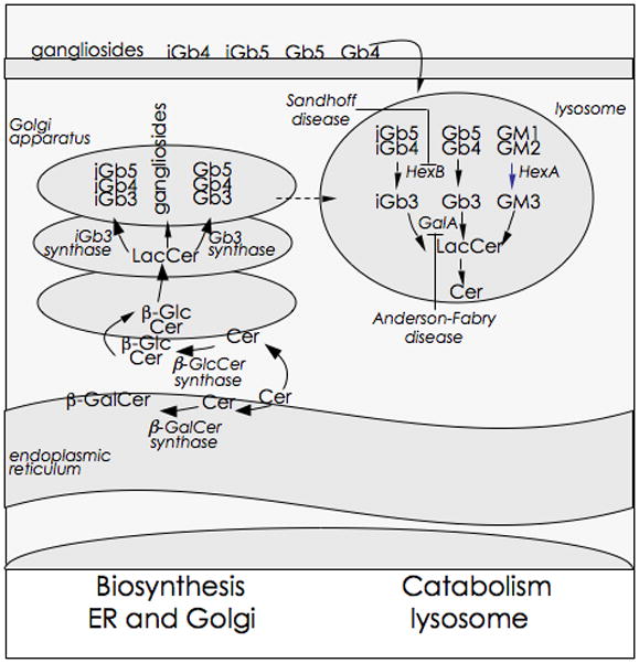
NKT cells that express the semi-invariant TCR are innate-like lymphocytes whose functions are regulated by self and foreign glycolipid ligands presented by the Ag-presenting, MHC class I-like molecule CD1d. Activation of NKT cells in vivo results in rapid release of copious amounts of effector cytokines and chemokines with which they regulate innate and adaptive immune responses to pathogens, certain types of cancers, and self-antigens. The nature of CD1d-restricted ligands, the manner in which they are recognized, and the unique effector functions of NKT cells suggest an immunoregulatory role for this T cell subset. Their ability to respond fast and our ability to steer NKT cell cytokine response to altered lipid ligands make them an important target for vaccine design and immunotherapies against autoimmune diseases. This review summarizes our current understanding of CD1d-restricted ligand recognition by NKT cells and how these innate-like lymphocytes regulate inflammation.


Figures

Figure 1
Figure 1. Cellular glycolipid gradients
Glycosphingolipid biosynthesis begins with the synthesis of ceramide on the cytosolic leaflet of the ER. The glycosylation of ceramides results in the formation of glycosphingolipids. The major precursors for mammalian glycosphingolipids are β-glucosylceramide (βGlcCer) and β-galactosylceramide (βGalCer). βGluCer is synthesised by βGlcCer synthase whose catalytic site is predisposed to the cytosolic side of the Golgi apparatus. In contrast, βGalCer is synthesised by an ER luminal enzyme βGalCer synthase. As shown, further glycosylation of βGlcCer results in lactosylceramide (LacCer), gangliosides, globosides and isoglobosides as well as lactoneo- and muco-series of glycolipids (not shown). These distribute to various membranous compartments including the lysosomes. The endogenous iNKT cell antigen iGb3 is generated by the enzymatic cleavage of β1–3GalNAc from iGb4 by β-hexosaminidase B (HexB). iGb3 appear to be rapidly catabolised to LacCer by the action of another lysosomal hydrolase, β-galactosidase A (GalA). Microbes, such as Salmonella, and derived products down regulate cellular GalA gene expression, which prevents the catabolism of the NKT cell agonist, iGb3, amongst other glycosphingolipids. Deficiencies in HexB and GalA are know to cause Sandhoff’s and Anderson-Fabry diseases, respectively. These lipid storage diseases impact such fundamental processes as macro-autophagy, mitochondrial function as well as protein and lipid trafficking and thereby alter cellular homeostasis. Put together, cellular lipid homeostasis regulates NKT cell function, which in turn can control inflammation.
Figure 2
Figure 2. Chemical structure and orientation of ligands in the mCD1d ABG
Top, chemical structures of the ligands; gray, portions not ordered in the corresponding crystal structures. Bottom, a side view of the ABG with the α2 helix removed for clarity. Ligand, yellow; spacer lipids, green; mouse CD1d heavy chain, gray; unsaturations on the acyl chains of the ligands are also green. Some of the residues involved in defining the ABG and contacting the ligand are highlighted. PDB ID: αGalCer (human), 1ZT4;αGalCer, 1Z5L; C6Ph, 3GML; OCH, 3G08; αGalACer, 2FIK; iGb3, 2Q7Y; sulfatide, 2AKR; αGalDAG, 3ILQ; PtdInoMan2, 2GAZ; PtdCho, 1ZHN.
Figure 3
Figure 3. Top view of the CD1d-ligand structures
A top view of the ABG is shown before NKTCR binding with the protein surface in grey and the ligands in yellow. Note the presence of the pre-formed F′ roof exclusively for αGalCer.
Figure 4
Figure 4. NKTCR/CD1d-lipid interactions
(A) Structure of the mouse CD1d-αGalCer-NKTCR complex (3HE6). Mouse CD1d, grey; β2m, light blue; TCR α-chain, cyan; TCRβ-chain, orange; αGalCer, yellow. Note how the contacts between the NKTCR and the ligand or CD1d are dominated by the TCR α-chain. (B–J) Conserved interaction of the NKTCR with α-anomeric galactose-containing ligands and with the PtdIno self antigen. The ligands are shown in yellow with CD1d in grey and the TCR α-chain in cyan. Hydrogen bonds between the ligand and conserved residues on the NKTCR are shown as dashed blue lines. (F) Superposition of the mouse CD1d-αGalDAG structure before (blue) and after (ligand in yellow; CD1d, gray; TCR α-chain, cyan) TCR binding. Note how a conformational change of the galactose on the ligand is induced upon binding of the NKTCR α-chain in order to avoid steric clashes and allow for the conserved TCR binding footprint onto CD1d. PDB IDs: (AB) αGalCer, 3HE6; (C) αGalACer, 3O8X; (D)αGalCer (human), 3HUJ; (E) αGalDAG, 3O9W; (F) αGalDAG, 3O9W and 3ILQ; (G) PtdIno, 3QI9; (H) OCH, 3ARB; (I) α-C-GalCer, 3QUX; (J) NU-αGalCer, 3QUZ.

Similar articles

Cited by

References

    1. Medzhitov R. Origin and physiological roles of inflammation. Nature. 2008;454:428–435. - PubMed
    1. Bezbradica JS, Stanic AK, Matsuki N, Bour-Jordan H, Bluestone JA, Thomas JW, Unutmaz D, Van Kaer L, Joyce S. Distinct roles of dendritic cells and B cells in Va14Ja18 natural T cell activation in vivo. J Immunol. 2005;174:4696–4705. - PubMed
    1. Schmieg J, Yang G, Franck RW, Van Rooijen N, Tsuji M. Glycolipid presentation to natural killer T cells differs in an organ-dependent fashion. Proc Natl Acad Sci U S A. 2005;102:1127–1132. - PMC - PubMed
    1. Skold M, Xiong X, Illarionov PA, Besra GS, Behar SM. Interplay of cytokines and microbial signals in regulation of CD1d expression and NKT cell activation. J Immunol. 2005;175:3584–3593. - PubMed
    1. Winau F, Hegasy G, Weiskirchen R, Weber S, Cassan C, Sieling PA, Modlin RL, Liblau RS, Gressner AM, Kaufmann SH. Ito cells are liver-resident antigen-presenting cells for activating T cell responses. Immunity. 2007;26:117–129. - PubMed

Publication types

MeSH terms