. 2011 Jul 19;30(16):3285-97.
doi: 10.1038/emboj.2011.243.

Recognition of UbcH5c and the nucleosome by the Bmi1/Ring1b ubiquitin ligase complex

Affiliations

Recognition of UbcH5c and the nucleosome by the Bmi1/Ring1b ubiquitin ligase complex

Matthew L Bentley et al. EMBO J. .

Abstract

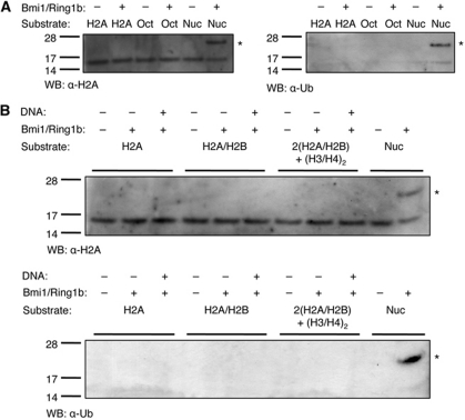
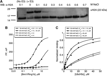
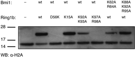
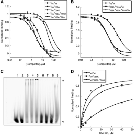
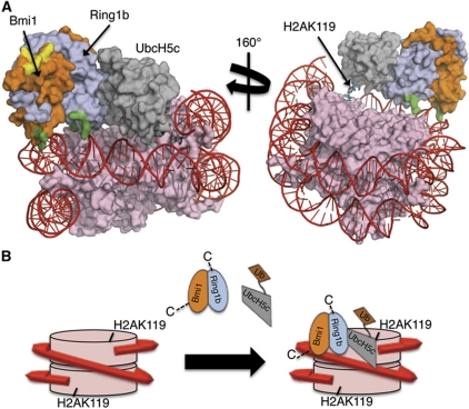
The Polycomb repressive complex 1 (PRC1) mediates gene silencing, in part by monoubiquitination of histone H2A on lysine 119 (uH2A). Bmi1 and Ring1b are critical components of PRC1 that heterodimerize via their N-terminal RING domains to form an active E3 ubiquitin ligase. We have determined the crystal structure of a complex between the Bmi1/Ring1b RING-RING heterodimer and the E2 enzyme UbcH5c and find that UbcH5c interacts with Ring1b only, in a manner fairly typical of E2-E3 interactions. However, we further show that the Bmi1/Ring1b RING domains bind directly to duplex DNA through a basic surface patch unique to the Bmi1/Ring1b RING-RING dimer. Mutation of residues on this interaction surface leads to a loss of H2A ubiquitination activity. Computational modelling of the interface between Bmi1/Ring1b-UbcH5c and the nucleosome suggests that Bmi1/Ring1b interacts with both nucleosomal DNA and an acidic patch on histone H4 to achieve specific monoubiquitination of H2A. Our results point to a novel mechanism of substrate recognition, and control of product formation, by Bmi1/Ring1b.


Conflict of interest statement

The authors declare that they have no conflict of interest.

Figures

Figure 1
Figure 1
Structure of the Bmi1/Ring1b–UbcH5c complex. (A) (Left) Ribbon representation of the overall complex architecture, with UbcH5c in grey, Ring1b(1−116) in light blue, and Bmi1(1−109) in orange. Zn2+ ions are shown in pink. The three Ring1b-binding regions on the surface of UbcH5c are coloured in teal (N-terminal α-helix), yellow (L4 loop), and purple (L7 loop), respectively. The sidechain of the catalytic Cys85 residue (site of ubiquitin attachment) is shown in green in stick format. (Right) Bmi1/Ring1b are shown in surface representation, highlighting the UbcH5c-binding groove on Ring1b. (B) Interactions between Ring1b and UbcH5c along the L4 and L7 loops (left), and N-terminal α-helix (right). The sidechains involved are shown in stick format. Hydrogen-bond distances are given in angstroms. Loops are coloured as in part (A). Key: nitrogen=blue, oxygen=red, sulphur=yellow, Zn2+=pink spheres, H2O=tan spheres.
Figure 2
Figure 2
Substrate preference of the Bmi1/Ring1b E3 ligase. (A) Reactions were incubated in ligase buffer containing 30 nM human E1, 1.5 μM UbcH5c, 25 μM ubiquitin, 3 mM ATP, 500 nM Bmi1(1−109)/Ring1b(1−116), and 5 μg of recombinant histone H2A (H2A), core histone octamers (Oct), or nucleosomes (Nuc). Reactions were incubated for 1 h at 30 °C, then separated by SDS–PAGE and analysed by western blot using either an anti-histone H2A (left) or anti-ubiquitin (right) antibody. Symbol: * represents ubiquitin H2A (22 kDa). (B) Added DNA is not sufficient to rescue uH2A activity towards non-nucleosomal substrates. Recombinant histone H2A (H2A), recombinant H2A/H2B dimer (H2A/H2B), 2:1 H2A/H2B dimer:H3/H4 tetramer (2(H2A/H2B)+(H3/H4)2), or nucleosomes were treated in the presence or absence of equimolar concentrations of a 146-bp DNA duplex. All reactions contained 2.5 μM histone H2A, and all other conditions were the same as in (A). Symbol: * represents ubiquitin H2A (22 kDa).
Figure 3
Figure 3
Bmi1/Ring1b binds to short duplex DNA. (A, B) In all, 70 nM 5′-FAM-labelled DNA probe was incubated with indicated concentrations of Bmi1/Ring1b at room temperature for 10 min. Polarization data are plotted as average values±s.d. at each concentration. (A) Direct binding assay, showing increase in FP as a function of [Bmi1/Ring1b] KD,app=3.2±0.3 μM. (B) Competition binding assay. Bmi1/Ring1b (2.5 μM) was mixed with 70 nM probe, and unlabelled DNA (same sequence as probe) was used to compete for binding. IC50=3.0±1.0 μM. (C) DNA binding is non-sequence specific. Four scrambled sequences (see Table II) were tested in the competition assay as in (B). (D) Length dependence of DNA binding. 3′ truncations of the original 12 bp sequence were assayed as in (B). In (C, D), data were normalized and plotted as average values±s.d. IC50 values were determined from fitting the average values to a four-parameter fit.
Figure 4
Figure 4
Salt dependence of Bmi1/Ring1b ubiquitin ligase activity, UbcH5c binding, and DNA binding. (A) Histone H2A ubiquitin ligase assay in the presence of added salt. Bmi1/Ring1b was incubated in ligase buffer with nucleosomes under the conditions described in Figure 2, with [NaCl] allowed to vary from 0.1 to 0.7 M. H2A ubiquitin ligase activity is highly salt dependent, with complete loss of product formation at 0.3 M NaCl. (B) DNA binding as a function of NaCl concentration. The FP assay was conducted as described in Figure 3A at each indicated [NaCl]. (C) UbcH5c binding as a function of added NaCl. Immobilized Bmi1/Ring1b was exposed to increasing concentrations of UbcH5c, and binding was detected by BLI. Normalized steady-state responses at equilibrium are plotted as a function of UbcH5c concentration, and KD values were determined as described in Materials and methods.
Figure 5
Figure 5
Ubiquitin ligase activity of Bmi1/Ring1b mutants. The indicated mutants (Bmi1/Ring1b heterodimers) were incubated in ligase buffer (1 h; 30°C) with E1, UbcH5c, ubiquitin, ATP, and nucleosomes. Products were analysed as described above.
Figure 6
Figure 6
Mutation of basic surface residues on Bmi1/Ring1b disrupts DNA binding without affecting UbcH5c binding. (A, B) FP DNA-binding assay of Bmi1/Ring1b mutants. Binding of heterodimers bearing mutations in Ring1b is shown in (A), and in (B) for those bearing mutations in Bmi1. Complex (at a concentration that gave rise to a 50-mP change in signal from a free probe baseline) was incubated with 70 nM 5′-FAM-labelled probe, and unlabelled probe was used to compete for binding. Raw mP values were converted to normalized binding curves. Data are plotted as average values±s.d. (C) F-EMSA assay of Bmi1/Ring1b mutants. In all, 20 μM wt or mutant protein was incubated with 70 nM 5′-FAM probe for 10 min at room temperature before electrophoretic separation. Symbols: * represents Bmi1/Ring1b–probe complex and ° represents free probe. Lane key: 1=blank (H2O), 2=blank (buffer), 3=Bwt/Rwt, 4=Bwt/RD56K, 5=Bwt/RK15A, 6=Bwt/RK92A.RK93A, 7=Bwt/RK97A.RR98A, 8=BK62A.BR64A/Rwt, 9=BK88A.BK92A.BR95A/Rwt. (D) BLI (Octet)-binding assay of UbcH5c to Bwt/Rwt, Bwt/RD56K, or Bwt/RK97A.R98A. Normalized responses (binding) are plotted as a function of UbcH5c concentration, and KD values were determined as described in Materials and methods.
Figure 7
Figure 7
Model for the interaction of the Bmi1/Ring1b–UbcH5c complex with the nucleosome. (A) Computational docking model generated using HADDOCK2.0. Colour coding is as follows: histones=pink, DNA=red, Ring1b=light blue, Bmi1=orange, UbcH5c=grey. Basic surface residues of Bmi1/Ring1b that were mutated in this study are shaded according to their effect on DNA binding: green=mutation that affected DNA binding, yellow=mutation that had no effect on DNA binding. The C-terminus of histone H2A (cyan stick representation) is the site of ubiquitin modification (K119). UCys85 is coloured by element: nitrogen=blue, oxygen=red, sulphur=yellow. (B) Cartoon model for recognition of the nucleosome by Bmi1/Ring1b–UbcH5c∼Ub complex. Colour scheme is the same as above (ubiquitin is shown in brown). Bmi1/Ring1b uses the basic saddle region to recognize and dock onto the nucleosome through contacts to both DNA and histone H4, positioning UbcH5c∼Ub for ubiquitin transfer to H2AK119. Following transfer of a single ubiquitin, UbcH5c∼Ub is no longer able to access both the E2-binding site on Ring1b and the nucleosome at the same time, leading to termination of the cycle after a single ubiquitination event.

References

    1. Adams PD, Afonine PV, Bunkoczi G, Chen VB, Davis IW, Echols N, Headd JJ, Hung LW, Kapral GJ, Grosse-Kunstleve RW, McCoy AJ, Moriarty NW, Oeffner R, Read RJ, Richardson DC, Richardson JS, Terwilliger TC, Zwart PH (2010) PHENIX: a comprehensive Python-based system for macromolecular structure solution. Acta Crystallogr 66(Part 2): 213–221 - PMC - PubMed
    1. Ben-Saadon R, Zaaroor D, Ziv T, Ciechanover A (2006) The polycomb protein Ring1B generates self atypical mixed ubiquitin chains required for its in vitro histone H2A ligase activity. Mol Cell 24: 701–711 - PubMed
    1. Bernstein E, Duncan EM, Masui O, Gil J, Heard E, Allis CD (2006) Mouse polycomb proteins bind differentially to methylated histone H3 and RNA and are enriched in facultative heterochromatin. Mol Cell Biol 26: 2560–2569 - PMC - PubMed
    1. Bracken AP, Helin K (2009) Polycomb group proteins: navigators of lineage pathways led astray in cancer. Nat Rev Cancer 9: 773–784 - PubMed
    1. Bruggeman SW, Valk-Lingbeek ME, van der Stoop PP, Jacobs JJ, Kieboom K, Tanger E, Hulsman D, Leung C, Arsenijevic Y, Marino S, van Lohuizen M (2005) Ink4a and Arf differentially affect cell proliferation and neural stem cell self-renewal in Bmi1-deficient mice. Genes Dev 19: 1438–1443 - PMC - PubMed

MeSH terms

Associated data