. 2011 Oct;19(10):1905-12.
doi: 10.1038/mt.2011.135. Epub 2011 Jul 19.

Rapid and efficient generation of functional motor neurons from human pluripotent stem cells using gene delivered transcription factor codes

Affiliations

Rapid and efficient generation of functional motor neurons from human pluripotent stem cells using gene delivered transcription factor codes

Mark E Hester et al. Mol Ther. 2011 Oct.

Abstract

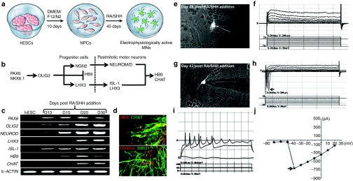
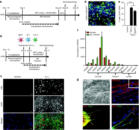
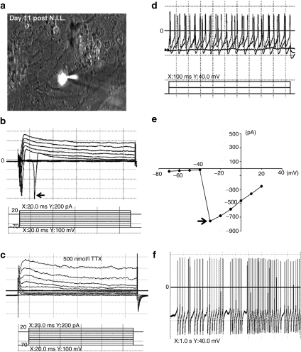
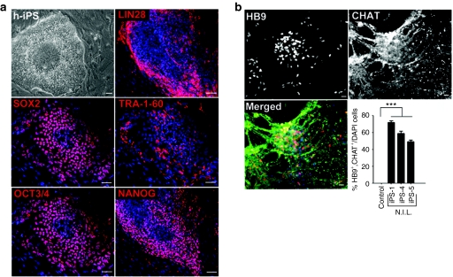
Stem cell-derived motor neurons (MNs) are increasingly utilized for modeling disease in vitro and for developing cellular replacement strategies for spinal cord injury and diseases such as spinal muscular atrophy (SMA) and amyotrophic lateral sclerosis (ALS). Human embryonic stem cell (hESC) differentiation into MNs, which involves retinoic acid (RA) and activation of the sonic hedgehog (SHH) pathway is inefficient and requires up to 60 days to develop MNs with electrophysiological properties. This prolonged differentiation process has hampered the use of hESCs, in particular for high-throughput screening. We evaluated the MN gene expression profile of RA/SHH-differentiated hESCs to identify rate-limiting factors involved in MN development. Based on this analysis, we developed an adenoviral gene delivery system encoding for MN inducing transcription factors: neurogenin 2 (Ngn2), islet-1 (Isl-1), and LIM/homeobox protein 3 (Lhx3). Strikingly, delivery of these factors induced functional MNs with mature electrophysiological properties, 11-days after gene delivery, with >60-70% efficiency from hESCs and human induced pluripotent stem cells (hiPSCs). This directed programming approach significantly reduces the time required to generate electrophysiologically-active MNs by approximately 30 days in comparison to conventional differentiation techniques. Our results further exemplify the potential to use transcriptional coding for rapid and efficient production of defined cell types from hESCs and hiPSCs.


Figures

Figure 1
Figure 1
hESCs require a long differentiation and maturation period to differentiate into functional MNs in vitro. (a) Differentiation paradigm of hESCs into MNs. hESCs were differentiated into neural progenitor cells in DMEM/F12/N2 media followed by RA/SHH treatment to induce MN differentiation. (b) Schematic depicting critical signaling factors in MN development. (c) RT-PCR analysis of markers involved in early and late MN differentiation assayed at multiple time points post RA/SHH treatment. (d) Mature MNs coexpress HB9 (red) and CHAT (green), and also express SMI31 (green) and express a marker of cervical spinal cord identity, HOXC6 (red). (e) Patch-clamped Hb9::RFP MN at 28 days post RA/SHH addition showed no sodium current activity (f). (g) Patch-clamped Hb9::RFP MN 2 weeks later showed sodium current activity (h), action potentials (i), and peak sodium current activity of approximately −700 pA (j). Arrow in panel “h” shows peaks representing sodium currents. Arrow in panel “j” shows the peak sodium current activity. In panel “d” bar = 50 µm. CHAT, choline acetyltransferase; DMEM, Dulbecco's modified Eagle's medium; hESCs, human embryonic stem cells; MN, motor neurons; RA, retinoic acid; RT-PCR, reverse transcriptase-PCR; SHH, sonic hedgehog.
Figure 2
Figure 2
Rapid and efficient MN differentiation of hESCs by the N.I.L. transcription factors. (a) Time line of differentiating human ES or iPS cells towards electrophysiologically-competent MNs utilizing RA/SHH. (b) Time line of accelerating human ES or iPS cell differentiation towards electrophysiologically-competent MNs utilizing N.I.L. (c) N.I.L.-induced MN differentiation showed co-expression of mature MN markers HB9 and CHAT. (d) HuES-3 Hb9::GFP cells differentiated into MNs with the N.I.L. transcription factor code show GFP expression. (e) Quantification of colabeled HB9/CHAT induced MNs. (f) qRT-PCR analysis of HOX genes expressed in the cervical, thoracic, and lumbar regions of the spinal cord assayed from N.I.L.-induced MNs and RA/SHH-induced MNs. (g) Phase contrast picture of C2C12-derived myotubes, which express α-actinin, with a high-power insert showing characteristic striations. (g) N.I.L.-induced MNs express SV2 (red) and colocalize with axons labeled with TUJ1 (green). MN axon terminals form junctions with acetylcholine receptors labeled with bungarotoxin (Red) as shown by an arrow (bottom panel). Arrow in panel “g” shows the colocalization of a MN axon with acetylcholine receptors on a myotube. In panel “c” all bars = 50 µm. In panel “g” all bars = 20 µm. DMEM, Dulbecco's modified Eagle's medium; hESCs, human embryonic stem cells; MN, motor neurons; N.I.L., Ngn2, Isl-1, and Lhx3; RA, retinoic acid; qRT-PCR, quantitative reverse transcriptase-PCR; SHH, sonic hedgehog.
Figure 3
Figure 3
N.I.L.-induced MNs are functionally mature showing characteristic electrophysiological properties. (a) N.I.L.-induced MN labeled with Hb9::RFP and patch clamped showing sodium current activity (b) that could be blocked with tetrodotoxin (TTX) (c). N.I.L.-induced MNs displayed action potentials (d), peak sodium current activity of approximately −800 pA (e), and spontaneous activity (f). Arrow in panel “b” shows peaks representing sodium currents. Arrow in panel “e” shows the peak sodium current activity. MN, motor neurons; N.I.L., Ngn2, Isl-1, and Lhx3.
Figure 4
Figure 4
N.I.L.-induces rapid and efficient generation of MNs from hiPSCs. (a) Phase contrast picture of a hiPSC clone showing distinct hESC morphology. hiPSCs express markers associated with pluripotency such as LIN28, OCT3/4, SOX2, TRA-1-60, and NANOG. (b) N.I.L.-induced MNs express HB9 and CHAT, and are colocalized in the same cells merged with the nuclear dye, DAPI. Quantification of induced MNs colabeled with HB9/CHAT from hiPS cells. In panel “a” bars = 100 µm. In panel “b” bars = 50 µm. hESC, human embryonic stem cell; hiPSCs, human induced pluripotent stem cells; MN, motor neurons; N.I.L., Ngn2, Isl-1, and Lhx3.

References

    1. Foust KD, Wang X, McGovern VL, Braun L, Bevan AK, Haidet AM.et al. (2010Rescue of the spinal muscular atrophy phenotype in a mouse model by early postnatal delivery of SMN Nat Biotechnol 28271–274. - PMC - PubMed
    1. Dominguez E, Marais T, Chatauret N, Benkhelifa-Ziyyat S, Duque S, Ravassard P.et al. (2011Intravenous scAAV9 delivery of a codon-optimized SMN1 sequence rescues SMA mice Hum Mol Genet 20681–693. - PubMed
    1. Passini MA, Bu J, Roskelley EM, Richards AM, Sardi SP, O'Riordan CR.et al. (2010CNS-targeted gene therapy improves survival and motor function in a mouse model of spinal muscular atrophy J Clin Invest 1201253–1264. - PMC - PubMed
    1. Valori CF, Ning K, Wyles M, Mead RJ, Grierson AJ, Shaw PJ.et al. (2010Systemic delivery of scAAV9 expressing SMN prolongs survival in a model of spinal muscular atrophy Sci Transl Med 235ra42 - PubMed
    1. Ebert AD, Yu J, Rose FF, Jr, Mattis VB, Lorson CL, Thomson JA.et al. (2009Induced pluripotent stem cells from a spinal muscular atrophy patient Nature 457277–280. - PMC - PubMed

Publication types

MeSH terms

Substances