

Endoscopic ultrasonography for gastric submucosal lesions
- PMID: 21772939
- PMCID: PMC3139278
- DOI: 10.4253/wjge.v3.i5.86
Endoscopic ultrasonography for gastric submucosal lesions
Abstract
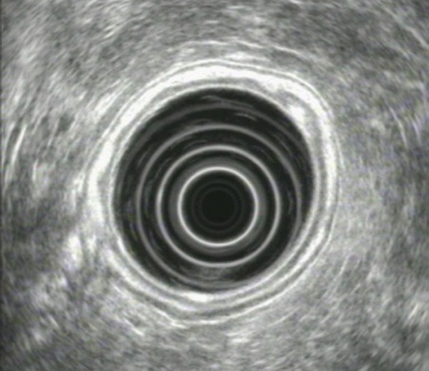
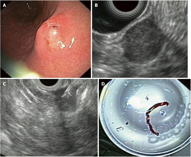
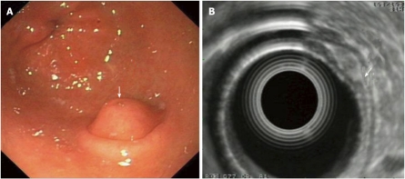
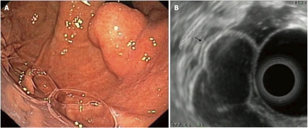
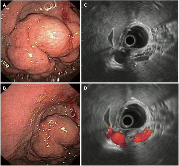
Gastric submucosal tumors (SMTs) are a rather frequent finding, occurring in about 0.36% of routine upper GI-endoscopies. EUS has emerged as a reliable investigative procedure for evaluation of these lesions. Diagnostic Endoscopic ultrasonography (EUS) has the ability to differentiate intramural tumors from extraluminal compressions and can also show the layer of origin of gastric SMTs. Tumors can be further characterized by their layer of origin, echo pattern and margin. EUS-risk criteria of their malignant potential are presented, although the emergence of EUS-guided fne needle aspiration (EUS-FNA) has opened new indications for transmural tissue diagnosis and expanded the possibilities of EUS in SMTs of the stomach. Tissue diagnosis should address whether the SMT is a Gastrointestinal stromal tumour (GIST) or another tumor type and evaluate the malignant potential of a given GIST. However, there seems to be a lack of data on the optimal strategy in SMTs suspected to be GISTs with a negative EUS-FNA tissue diagnosis. The current management strategies, as well as open questions regarding their treatment are also presented.
Keywords: EUS-guided fne needle aspiration; Endoscopic ultrasound; Gastric submucosal tumors; Gastrointestinal stromal tumours.
Figures
References
-
- Röesch T. Endoscopic ultrasonography: equipment and technique. Gastrointest Endosc Clin N Am. 2005;15:13–31, vii. - PubMed
-
- Papanikolaou IS, Fockens P, Hawes R, Rösch T. Update on endoscopic ultrasound: how much for imaging, needling, or therapy? Scand J Gastroenterol. 2008;43:1416–1424. - PubMed
-
- Anderson MA, Scheiman JM. Initial experience with an electronic radial array echoendoscope: randomized comparison with a mechanical sector scanning echoendoscope in humans. Gastrointest Endosc. 2002;56:573–577. - PubMed
-
- Noh KW, Woodward TA, Raimondo M, Savoy AD, Pungpapong S, Hardee JD, Wallace MB. Changing trends in endosonography: linear imaging and tissue are increasingly the issue. Dig Dis Sci. 2007;52:1014–1018. - PubMed
-
- Rösch T. The radial echoendoscope: here to stay or gone tomorrow? Gastrointest Endosc. 2009;69:S159–S162. - PubMed
LinkOut - more resources
Full Text Sources
Medical
