

Evaluation of Biomarkers of NAFLD in a Cohort of Morbidly Obese Patients
- PMID: 21773018
- PMCID: PMC3135053
- DOI: 10.1155/2011/369168
Evaluation of Biomarkers of NAFLD in a Cohort of Morbidly Obese Patients
Abstract
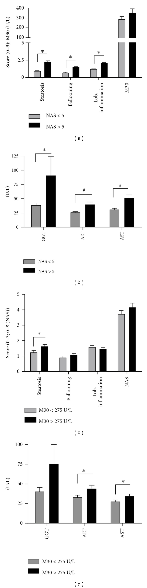
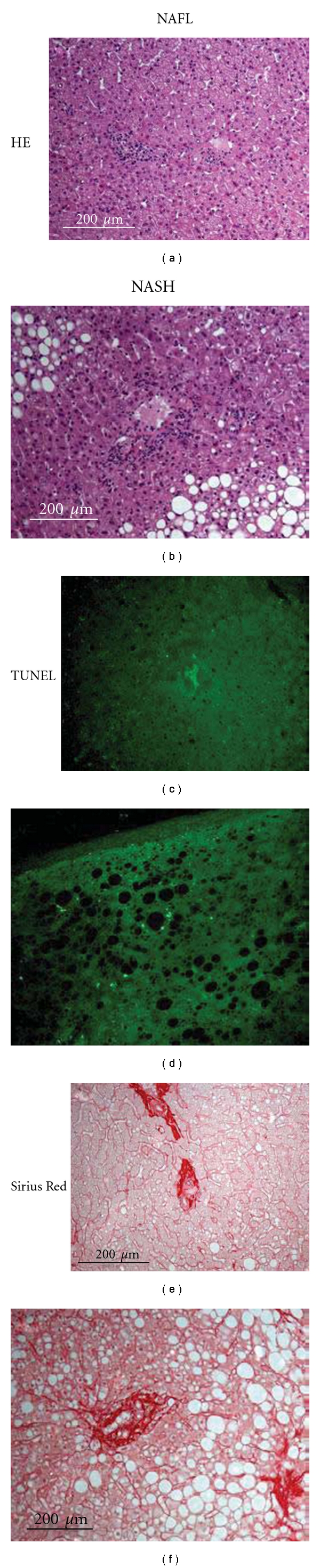
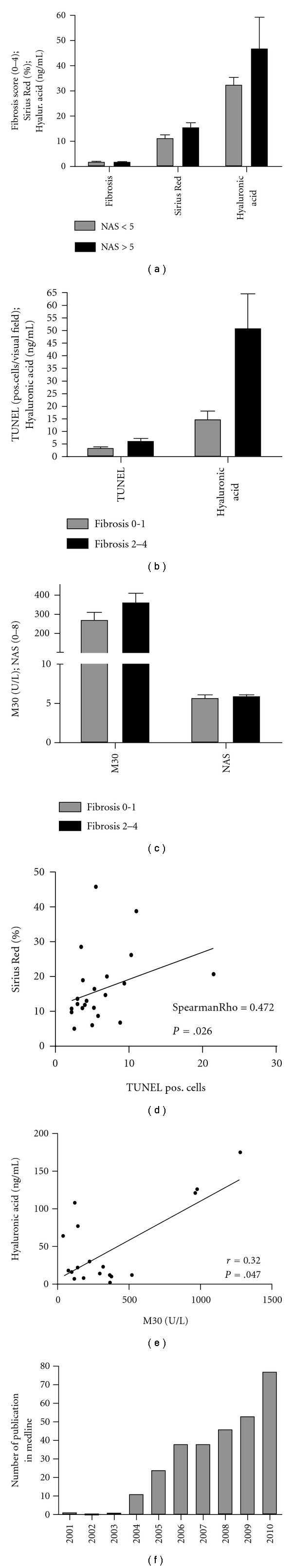
Hepatocyte apoptosis is a key event in nonalcoholic fatty liver disease (NAFLD), and serum apoptotic markers are emerging as surrogate markers for NAFLD. We studied the role of caspase-cleaved cytokeratin18 in the diagnosis of fibrosis in a cohort of 127 morbidly obese patients and also performed a review of the literature biomarkers of NAFLD and fibrosis. Here, we found that cleaved caspase 18 correlated with liver steatosis and liver injury as assessed by serum transaminase levels. Furthermore, hepatocyte apoptosis as assessed by cleaved CK18 and TUNEL staining correlated with the extent of fibrosis as assessed by Sirius Red staining and serum hyaluronic acid. These results underscore the important role of hepatocyte apoptosis in the pathogenesis of fibrosis in NAFLD, which led to the utilization of surrogate markers for apoptosis in the noninvasive diagnosis of NAFLD. We furthermore reviewed current literature of biomarkers of NAFLD and fibrosis.
Figures
References
-
- Feldstein AE. Novel insights into the pathophysiology of nonalcoholic fatty liver disease. Seminars in Liver Disease. 2010;30(4):391–401. - PubMed
-
- Cheung O, Sanyal AJ. Abnormalities of lipid metabolism in nonalcoholic fatty liver disease. Seminars in Liver Disease. 2008;28(4):351–359. - PubMed
-
- Ertle J, Dechêne A, Sowa JP, et al. Non-alcoholic fatty liver disease progresses to hepatocellular carcinoma in the absence of apparent cirrhosis. International Journal of Cancer. 2011;128(10):2436–2443. - PubMed
-
- Feldstein AE, Canbay A, Angulo P, et al. Hepatocyte apoptosis and fas expression are prominent features of human nonalcoholic steatohepatitis. Gastroenterology. 2003;125(2):437–443. - PubMed
-
- Wieckowska A, Zein NN, Yerian LM, Lopez AR, McCullough AJ, Feldstein AE. In vivo assessment of liver cell apoptosis as a novel biomarker of disease severity in nonalcoholic fatty liver disease. Hepatology. 2006;44(1):27–33. - PubMed
LinkOut - more resources
Full Text Sources
