. 2011 Oct;32(29):7106-17.
doi: 10.1016/j.biomaterials.2011.06.021. Epub 2011 Jul 20.

Discontinuities in the human bone-PDL-cementum complex

Affiliations

Discontinuities in the human bone-PDL-cementum complex

Jonathan M Hurng et al. Biomaterials. 2011 Oct.

Abstract

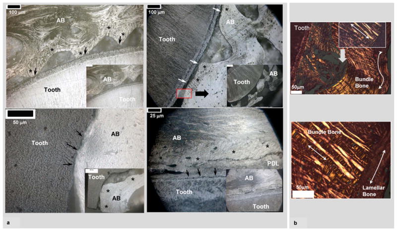
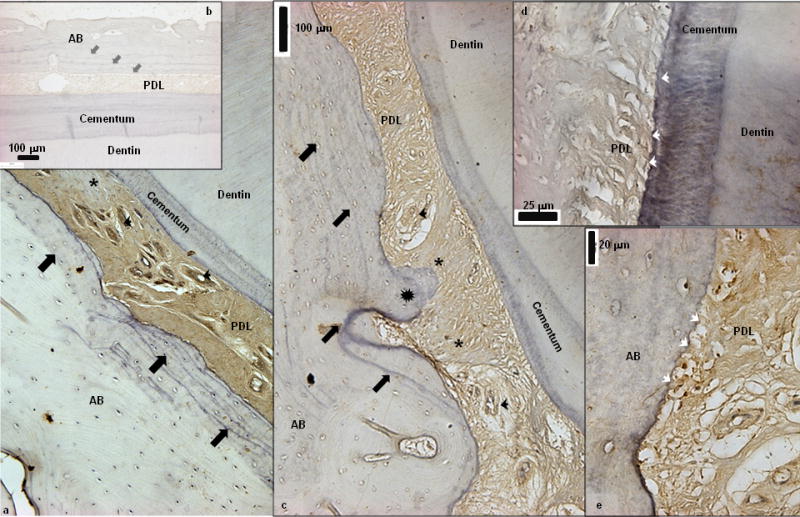
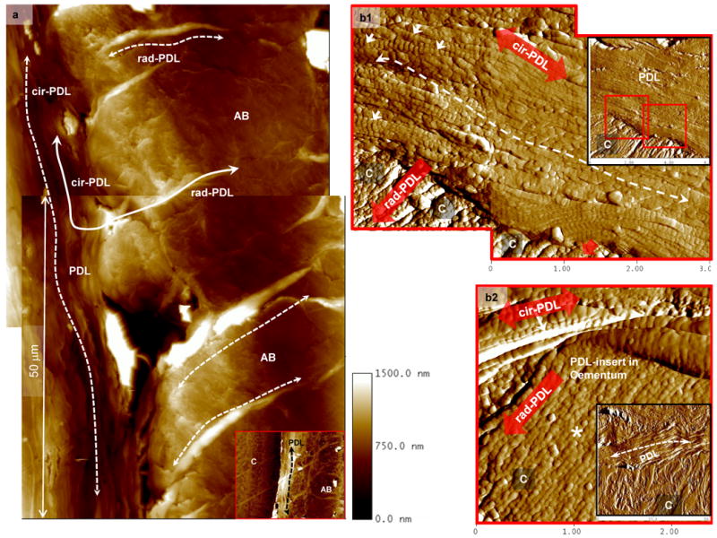
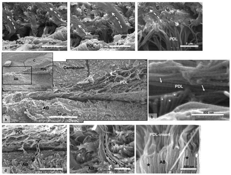
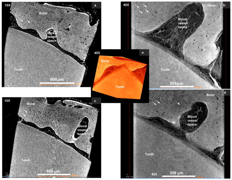
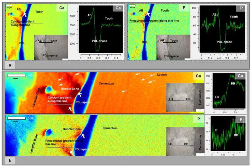
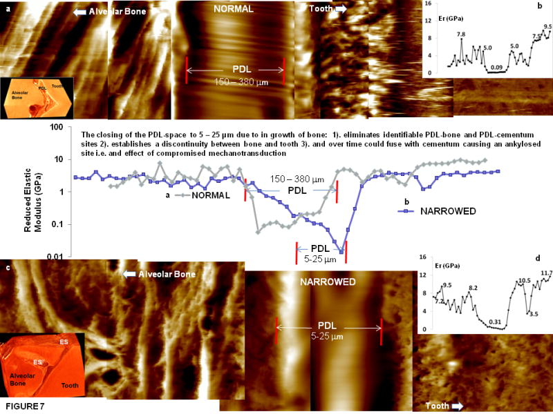
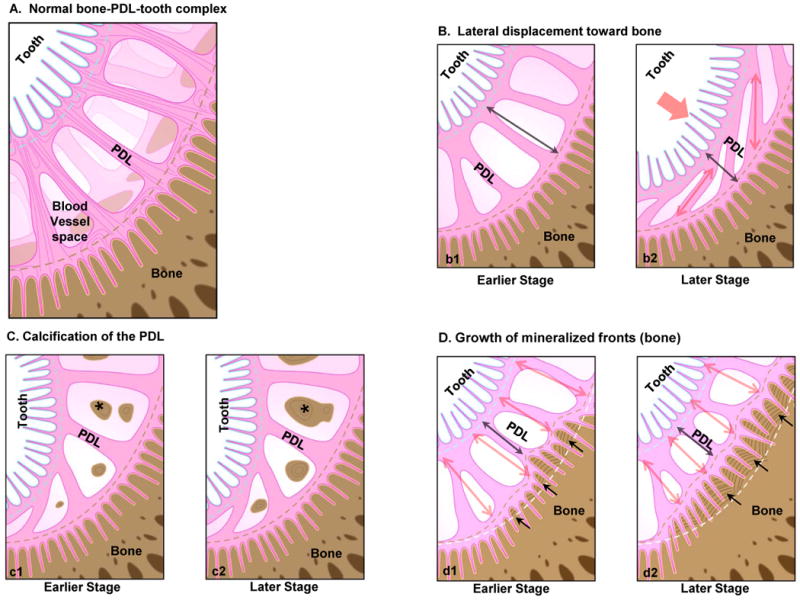
A naturally graded interface due to functional demands can deviate toward a discontinuous interface, eventually decreasing the functional efficiency of a dynamic joint. It is this characteristic feature in a human bone-tooth fibrous joint bone-PDL-tooth complex that will be discussed through histochemistry, and site-specific high resolution microscopy, micro tomography(Micro XCT™), X-ray fluorescence imaging and wet nanoindentation techniques. Results demonstrated two causes for the occurrence of 5-50 μm narrowed PDL-space: 1) microscopic scalloped regions at the PDL-insertion sites and macro-scale stratified layers of bone with rich basophilic lines, and 2) macroscopic bony protrusions. Narrowed PDL-complexes illustrated patchy appearance of asporin, and when imaged under wet conditions using an atomic force microscope (AFM), demonstrated structural reorganization of the PDL, collagen periodicity, organic-dominant areas at the PDL-cementum and PDL-bone entheses and within cementum and bone. Scanning electron microscopy (SEM) results confirmed AFM results. Despite the narrowed PDL, continuity between PDL and vasculature in endosteal spaces of bone was demonstrated using a Micro XCT™. The higher levels of Ca and P X-ray fluorescence using a microprobe were correlated with higher elastic modulus values of 0.1-1.4 and 0.1-1.2 GPa for PDL-bone and PDL-cementum using wet nanoindentation. The ranges in elastic modulus values for PDL-bone and PDL-cementum entheses in 150-380 μm wide PDL-complex were 0.1-1.0 and 0.1-0.6 GPa. Based on these results we propose that strain amplification at the entheses could be minimized with a gradual change in modulus profile, a characteristic of 150-380 μm wide functional PDL-space. However, a discontinuity in modulus profile, a characteristic of 5-50 μm wide narrowed PDL-space would cause compromised mechanotransduction. The constrictions or narrowed sites within the bone-tooth fibrous joint will become the new "load bearing sites" that eventually could cause direct local fusion of bone with cementum.


Figures

Figure 1
Figure 1
a) Low (insets) and high resolution light micrographs of ultrasectioned surface blocks illustrated narrowed PDL-space (arrows) between alveolar bone (AB) and tooth. All micrographs illustrate AB, which is a combination of bundle bone (asterisks) and lamellar bone. Bundle bone is significantly closer to cementum (C). b) Hematoxylin and eosin staining (H&E) of AB coupled with polarized light microscopy demonstrated dominant collagen fibers of different dimension and orientation within bundle bone (mainly radial fibers, straight arrows) compared to lamellar bone (mainly circumferential fibers, curved arrows). It should be noted that both types of bone within AB contain interwoven fabric-like structure.
Figure 2
Figure 2
a) Immunohistochemistry illustrated localization of asporin within PDL-space including vascular network (black arrow heads). Interestingly patchy staining indicative of heterogeneous distribution of asporin within the complex was observed (asterisks). Additionally, several basophilic lines representative of bone advancement into PDL-space can be observed (black block arrows) b) Negative control is shown as an inset. c) Stratified basophilic lines (lamellae, block arrows) in addition to directional bone growth (protrusions indicated by star burst into PDL-space was observed. d, e) High resolution light micrographs of PDL-cementum and PDL-bone attachment sites illustrated multiple micro protrusions, i.e. “scalloped” micro regions (white arrow heads). AB: Alveolar bone, PDL: Periodontal ligament.
Figure 3
Figure 3
a). AFM scans of wet complex illustrated swelling of radial PDL-inserts within AB (rad-PDL, dashed curved arrows) in addition to parallel or circumferential orientation of periodontal ligament (cir-PDL) (dashed curved arrows) between cementum (C) and alveolar bone (AB) (inset in Fig. 3a). b). The 20 μm wide PDL (insets in Figs. 3b1 and 3b2) parallel to the cementum (C) surface (dashed curved arrow) contains collagen fibrils (arrows) identified by their characteristic periodicity. PDL is attached to cementum via rad-PDL. b1) Representative micrographs for cementum and bone illustrated presence (arrows) and absence of periodicity within PDL collagen fibrils and PDL-inserts. b2) Additionally, PDL-inserts in cementum contained collagen fibrils with beaded appearance and lacking periodicity (asterisk). In all images, the insets are lower magnification AFM micrographs containing the highlighted regions of interest at higher magnifications.
Figure 4
Figure 4
a) SEM micrographs of cryofractured surfaces illustrated narrowed PDL-space between cementum (C) and alveolar bone (AB). The collagen fibrils within the narrowed PDL are parallel to cementum surface. b) Characteristic periodicity of collagen fibrils within PDL can be noticed (straight arrows) in addition to out-of-plane collagen fibrils (asterisks). c) Top row of micrographs illustrate PDL-inserts into cementum (dashed-curved arrows), while d) the bottom row illustrates PDL-inserts (dashed-curved arrows) into alveolar bone (AB). Note that the collagen fibrils within the PDL-inserts lack periodicity.
Figure 5
Figure 5
a) Micro XCT™ slices of the bone-tooth complex illustrated narrowed PDL-space, PDL-orientation and association with alveolar bone and tooth (a, c) at a lower magnifications of 10X, (b, d) higher magnification, 40X. All images including the 3D image illustrate network of the PDL continuous with the blood vessel space in the alveolar bone. b and d) Virtual sections taken from the 3D images illustrate the 2D network of the bone-PDL-tooth complex, in addition to bundle bone identified by radial PDL-inserts (arrows). Note: Movies demonstrating virtual sectioning of the narrowed PDL-complex taken at 10X and 40X are also included as supplementary data.
Figure 6
Figure 6
a) Low resolution μ-XRF area maps of bone-PDL-tooth complex illustrated regions with higher counts of Ca and P in bone and closer to the PDL-space. Line maps illustrated steep gradients in Ca and P at the bone-PDL attachment site/interface. White bar represents 90 μm. Insets in the area maps represent mapped regions. Images were taken using a light microscope. b) High resolution μ-XRF maps illustrated Ca and P dominant regions within bundle bone relative to lamellar bone. Additionally, several lacunae in cementum and bone were identified (white arrows). Line maps illustrated gradients in Ca and P between bundle bone and lamellar bone. Additionally heterogeneity in Ca and P can be observed in bone and cementum within the complex. White bar represents 100 μm. BB: Bundle bone, LB: Lamellar bone, Ca: Calcium, P: Phosphorus.
Figure 7
Figure 7
Line profiles of reduced elastic modulus values with significant gradients at bone-PDL and cementum-PDL attachment sites of normal (a) and narrowed (c) wet bone-PDL-tooth complex. b, d) Insets illustrate linear elastic modulus profiles of normal (b) and narrowed (d) bone-tooth complex. Insets in the left hand corner illustrate representative tomographies of a normal and narrowed bone-tooth complex. PDL: periodontal ligament, ES: endosteal space, Er: reduced elastic modulus (GPa).
Figure 8
Figure 8
a) Schematic of a healthy bone-tooth complex illustrating a normal PDL-space. b-d) Schematics provide plausible scenarios that can be activated locally due to macroscale loads eventually narrowing PDL-space. Macroscale functional loads can cause the tooth to displace spatially toward the alveolar bone compressing the PDL-space (b) resulting in circumferential PDL fibers (b2). Functional loads can cause tilting of the root within the alveolar bone socket (c), altering hydrostatic pressure of PDL and blood vessels within PDL, thus stimulating cell differentiation, and expression of biomolecules as nucleators promoting PDL mineralization (asterisks). Functional loads can also cause local tension and compression sites that dominate events within PDL, and at the bone-PDL and cementum-PDL attachment sites. One such event is alveolar bone modeling due to pull-out forces at the tethered ends of the PDL causing vectorial growth of mineralization fronts into PDL-space (d1). Incremental layers represent stratified growth of bone and the scallop-like features at the PDL-bone attachment sites representing directional mineralization and advancement into PDL-space (d2). Although not show, similar events can occur in cementum at a slower rate, but can seen more in vascularized bone. Double headed red arrows represent tension within the PDL, while black arrows represent movement of the mineral front (red dashed line) into PDL-space. Note: Figures not drawn to scale.

References

    1. Suresh S. Graded materials for resistance to contact deformation and damage. Science. 2001;292(5526):2447–51. - PubMed
    1. Fratzl P. Biomimetic materials research: what can we really learn from nature's structural materials? J R Soc Interface. 2007;4:637–42. - PMC - PubMed
    1. Thompson DAW. On Growth and Form. Cambridge University Press; 1961.
    1. Carter DR, Beaupre GS. Skeletal Function and Form Mechanobiology of Skeletal Development, Aging, and Regeneration. Cambridge University Press; 2001.
    1. Genin GM, Kent A, Birman V, Wopenka B, Pasteris JD, Marquez PJ, et al. Functional Grading of Mineral and Collagen in the Attachment of Tendon to Bone. Biophys J. 2009;97(4):976–85. - PMC - PubMed

Publication types