

PreTCR and TCRγδ signal initiation in thymocyte progenitors does not require domains implicated in receptor oligomerization
- PMID: 21775286
- PMCID: PMC3475409
- DOI: 10.1126/scisignal.2001765
PreTCR and TCRγδ signal initiation in thymocyte progenitors does not require domains implicated in receptor oligomerization
Abstract
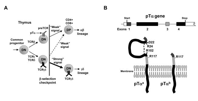
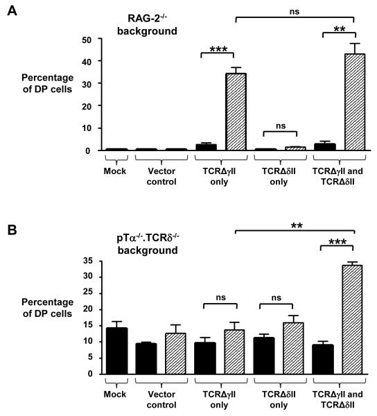
Whether thymocytes adopt an αβ or a γδ T cell fate in the thymus is determined at the β selection checkpoint by the relatively weak or strong signals that are delivered by either the pre-T cell receptor (preTCR) or the γδ TCR, respectively. Signal initiation at the β selection checkpoint is thought to be independent of ligand engagement of these receptors. Some reports have suggested that receptor oligomerization, which is thought to be mediated by either the immunoglobulin (Ig)-like domain of the preTCRα (pTα) chain or the variable domain of TCRδ, is a unifying mechanism that initiates signaling in early CD4(-)CD8(-) double-negative (DN) thymocyte progenitors. Here, we demonstrate that the extracellular regions of pTα and TCRδ that are implicated in mediating receptor oligomerization were not required for signal initiation from the preTCR or TCRγδ. Indeed, a truncated TCRγδ that lacked all of its extracellular Ig-like domains still formed a signaling-competent TCR that drove cells through the β selection checkpoint. These observations suggest that signal initiation in DN thymocytes is simply a consequence of the surface-pairing of TCR chains, with signal strength being a function of the abundances of surface TCRs. Thus, processes that regulate the surface abundances of TCR complexes in DN cells, such as oligomerization-induced endocytosis, would be predicted to have a major influence in determining whether cells adopt an αβ versus γδ T cell fate.
Figures
Similar articles
-
Human alpha beta and gamma delta thymocyte development: TCR gene rearrangements, intracellular TCR beta expression, and gamma delta developmental potential--differences between men and mice.J Immunol. 2006 Feb 1;176(3):1543-52. doi: 10.4049/jimmunol.176.3.1543. J Immunol. 2006. PMID: 16424183 Free PMC article.
-
Specific Notch receptor-ligand interactions control human TCR-αβ/γδ development by inducing differential Notch signal strength.J Exp Med. 2013 Apr 8;210(4):683-97. doi: 10.1084/jem.20121798. Epub 2013 Mar 25. J Exp Med. 2013. PMID: 23530123 Free PMC article. Clinical Trial.
-
Constitutive Notch signalling promotes CD4 CD8 thymocyte differentiation in the absence of the pre-TCR complex, by mimicking pre-TCR signals.Int Immunol. 2007 Dec;19(12):1421-30. doi: 10.1093/intimm/dxm113. Epub 2007 Nov 1. Int Immunol. 2007. PMID: 17981791
-
αβ versus γδ fate choice: counting the T-cell lineages at the branch point.Immunol Rev. 2010 Nov;238(1):169-81. doi: 10.1111/j.1600-065X.2010.00947.x. Immunol Rev. 2010. PMID: 20969592 Free PMC article. Review.
-
αβ and γδ T cell receptors: Similar but different.J Leukoc Biol. 2020 Jun;107(6):1045-1055. doi: 10.1002/JLB.2MR1219-233R. Epub 2020 Jan 29. J Leukoc Biol. 2020. PMID: 31994778 Review.
Cited by
-
Natural Self-Ligand Gamma Delta T Cell Receptors (γδTCRs) Insight: The Potential of Induced IgG.Vaccines (Basel). 2020 Aug 4;8(3):436. doi: 10.3390/vaccines8030436. Vaccines (Basel). 2020. PMID: 32759782 Free PMC article.
-
The TCR ligand-inducible expression of CD73 marks γδ lineage commitment and a metastable intermediate in effector specification.J Exp Med. 2014 Feb 10;211(2):329-43. doi: 10.1084/jem.20131540. Epub 2014 Feb 3. J Exp Med. 2014. PMID: 24493796 Free PMC article.
-
Developing T cells form an immunological synapse for passage through the β-selection checkpoint.J Cell Biol. 2021 Mar 1;220(3):e201908108. doi: 10.1083/jcb.201908108. J Cell Biol. 2021. PMID: 33464309 Free PMC article.
-
Diversity of IL-17-producing T lymphocytes.Cell Mol Life Sci. 2013 Jul;70(13):2271-90. doi: 10.1007/s00018-012-1163-6. Epub 2012 Oct 4. Cell Mol Life Sci. 2013. PMID: 23052209 Free PMC article. Review.
-
Developmental origins of murine γδ T-cell subsets.Immunology. 2019 Apr;156(4):299-304. doi: 10.1111/imm.13032. Epub 2019 Jan 21. Immunology. 2019. PMID: 30552818 Free PMC article. Review.
References
-
- Pennington DJ, Silva-Santos B, Hayday AC. Gammadelta T cell development--having the strength to get there. Curr Opin Immunol. 2005;17:108–115. - PubMed
-
- Hayday AC, Pennington DJ. Key factors in the organized chaos of early T cell development. Nat Immunol. 2007;8:137–144. - PubMed
-
- Mallick CA, Dudley EC, Viney JL, Owen MJ, Hayday AC. Rearrangement and diversity of T cell receptor beta chain genes in thymocytes: a critical role for the beta chain in development. Cell. 1993;73:513–519. - PubMed
-
- Fehling HJ, Krotkova A, Saint-Ruf C, von Boehmer H. Crucial role of the pre-T-cell receptor alpha gene in development of alpha beta but not gamma delta T cells. Nature. 1995;375:795–798. - PubMed
-
- Haks MC, Lefebvre JM, Lauritsen JP, Carleton M, Rhodes M, Miyazaki T, Kappes DJ, Wiest DL. Attenuation of gammadeltaTCR signaling efficiently diverts thymocytes to the alphabeta lineage. Immunity. 2005;22:595–606. - PubMed
Publication types
MeSH terms
Substances
Grants and funding
LinkOut - more resources
Full Text Sources
Molecular Biology Databases
Research Materials
