. 2011 Sep;138(17):3689-98.
doi: 10.1242/dev.057836. Epub 2011 Jul 20.

A Sox10 enhancer element common to the otic placode and neural crest is activated by tissue-specific paralogs

Affiliations

A Sox10 enhancer element common to the otic placode and neural crest is activated by tissue-specific paralogs

Paola Betancur et al. Development. 2011 Sep.

Abstract

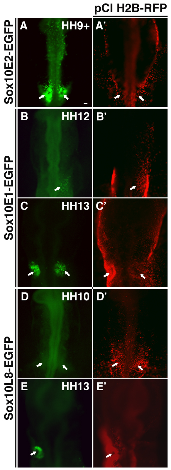
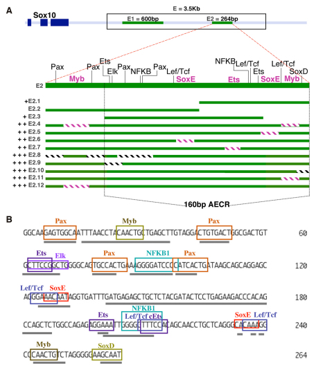
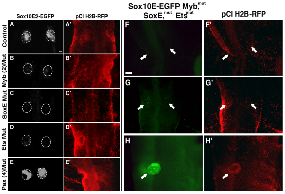
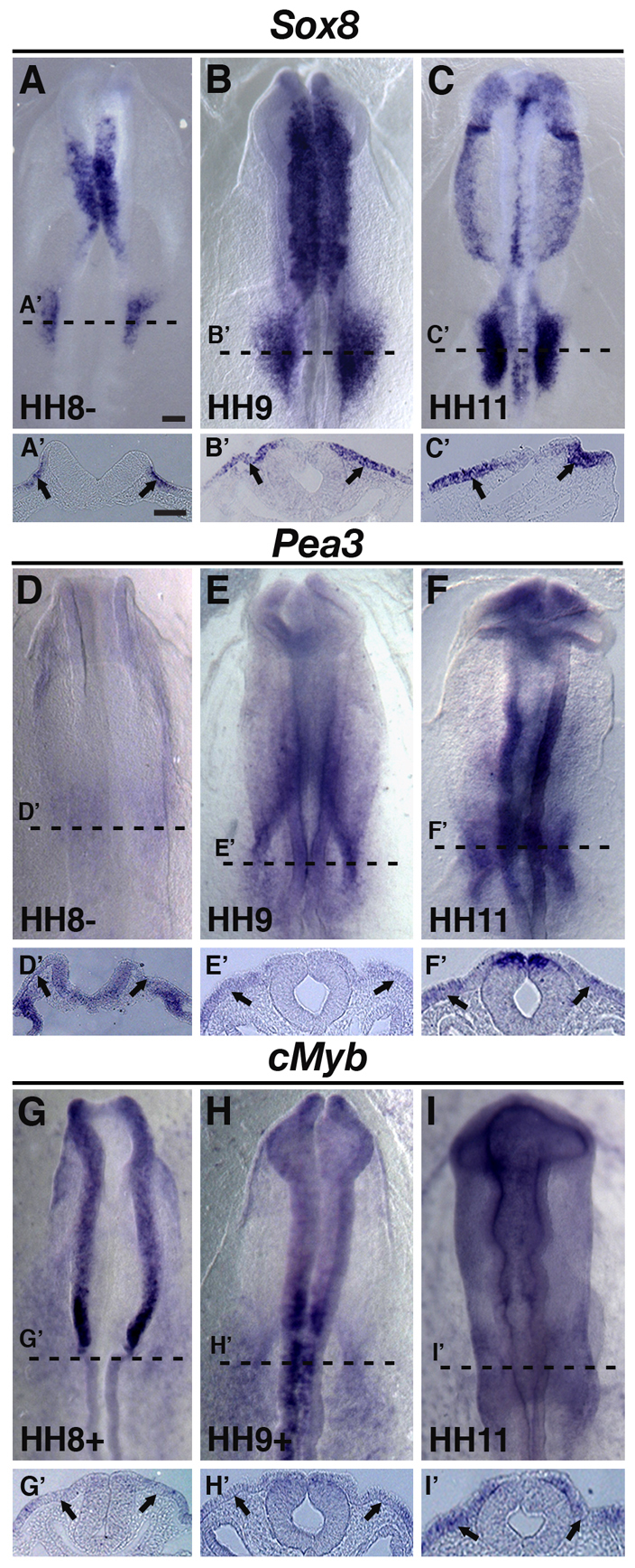
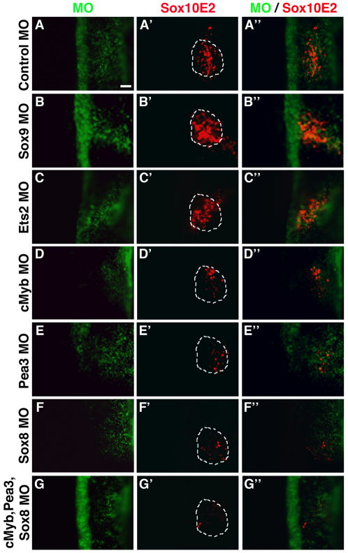
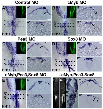
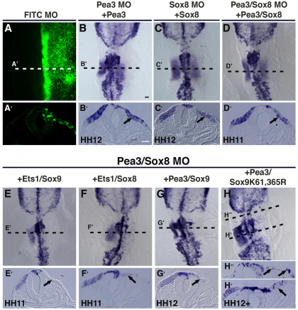
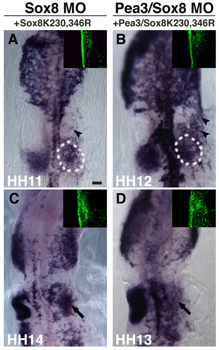
The otic placode, a specialized region of ectoderm, gives rise to components of the inner ear and shares many characteristics with the neural crest, including expression of the key transcription factor Sox10. Here, we show that in avian embryos, a highly conserved cranial neural crest enhancer, Sox10E2, also controls the onset of Sox10 expression in the otic placode. Interestingly, we show that different combinations of paralogous transcription factors (Sox8, Pea3 and cMyb versus Sox9, Ets1 and cMyb) are required to mediate Sox10E2 activity in the ear and neural crest, respectively. Mutating their binding motifs within Sox10E2 greatly reduces enhancer activity in the ear. Moreover, simultaneous knockdown of Sox8, Pea3 and cMyb eliminates not only the enhancer-driven reporter expression, but also the onset of endogenous Sox10 expression in the ear. Rescue experiments confirm that the specific combination of Myb together with Sox8 and Pea3 is responsible for the onset of Sox10 expression in the otic placode, as opposed to Myb plus Sox9 and Ets1 for neural crest Sox10 expression. Whereas SUMOylation of Sox8 is not required for the initial onset of Sox10 expression, it is necessary for later otic vesicle formation. This new role of Sox8, Pea3 and cMyb in controlling Sox10 expression via a common otic/neural crest enhancer suggests an evolutionarily conserved function for the combination of paralogous transcription factors in these tissues of distinct embryological origin.


Figures

Fig. 1.
Fig. 1.
Distinct EGFP reporter activity driven by the Sox10 enhancers Sox10E2, Sox10E1 and Sox10L8 is observed at different stages of otic development. (A-E′) EGFP reporter expression is activated by the Sox10 enhancer Sox10E2 as the otic placode becomes distinguishable at stage HH9+ in chicken embryos (A, arrows). By contrast, EGFP reporter expression under the control of enhancers Sox10E1 (B,C) and Sox10L8 (D,E) only appears later as the otic vesicle begins to invaginate (arrows). Panels A'-E' show the same embryos as in A-E, respectively, and show near ubiquitous expression of the co-electroporated tracer pCI H2B-RFP. Scale bar: 50 μm.
Fig. 2.
Fig. 2.
Analysis of reporter expression driven by different versions of enhancer Sox10E2 in the otic placode. (A) Diagram showing enhancers Sox10E1 and Sox10E2 located downstream of the dark blue squares representing Sox10 exons. Modified versions of Sox10E2 were created by dissecting the 264-bp fragment into smaller fragments, cutting within or around the 160-bp amniotic evolutionary conserved region (AECR; area between black dotted lines), or by mutating computationally located putative binding motifs (blocks of hatched lines). E2.8-E2.12 represent subfragments of Sox10E2 that showed strong regulatory activity in the otic placode. Sox10 E2.1 and E2.3-E2.7 subfragments displayed weak enhancing activity and E2.2 had no regulatory activity. +, ++ and +++ represent low, medium and high levels of observed reporter expression, respectively. Blocks of lavender hatched lines represent identified functional binding motifs. (B) Binding motifs for transcription factors identified computationally are depicted within the Sox10E2 genomic sequence. Gray underlines mark the specific nucleotide sequences that were mutated to test whether the individual or combination of binding motifs were relevant for the function of the Sox10E2 regulatory region.
Fig. 3.
Fig. 3.
SoxE, Ets and Myb binding motifs are required for the early enhancing activity of module Sox10E2 in the otic placode. (A-E) EGFP reporter expression is greatly decreased in the otic placode of HH11-HH12 chicken embryos after mutating both Myb sites (B), and either SoxE (C) or Ets (D) binding motifs within enhancer Sox10E2 compared with control (A). By contrast, no significant change in reporter expression is observed when mutating four Pax binding motifs simultaneously (E). Dotted outline demarcates the otic placode area. Scale bar: 50 μm. (F-H) Furthermore, a 3.5-kb Sox10E genomic fragment bearing mutations in SoxE, Ets and Myb binding motifs within the Sox10E2 enhancer region, but intact Sox10E1 enhancer, completely abolishes EGFP expression at HH9+ when endogenous Sox10 expression is first observed in the forming otic placode (F, arrows). Weak and scattered EGFP activity reappears around HH12-HH13 (G, arrows) when the otic vesicle begins to take shape and is back to normal levels around HH15 (H, arrow). Scale bar: 100 μm. A′-H′ correspond to A-H respectively and show efficient expression of the co-electroporated tracer pCI H2B-RFP.
Fig. 4.
Fig. 4.
Sox8, Pea3 and cMyb are expressed in the presumptive otic area prior to placode formation and Sox10 expression. (A-F′) By HH8, Sox8 and Pea3 transcripts are observed in the presumptive otic placode region (A,A',D,D' arrows). At HH9+ and HH11, Sox8 and Pea3 continue to be expressed in the developing otic placode (B-C',E-F', arrows). (G-I′) Endogenous cMyb is also observed at HH9-HH11 in the presumptive otic and forming placode region (arrows). A′-I′ show cross-sectional views of A-I, respectively, taken at the level shown by the dashed line. Scale bars: 100 μm for A-I; 50 μm for A'-I'.
Fig. 5.
Fig. 5.
cMyb, Pea3 or Sox8 morpholino-mediated knockdown dramatically reduces Sox10E2 regulatory activity in the otic placode. (A-A′) FITC-labeled morpholino control (green) does not affect Sox10E2-driven Cherry reporter expression (red) in the otic placode of HH11-HH12 chicken embryos. (B-C′) Similarly, Sox9 (B-B′) or Ets2 (C-C′) morpholinos do not affect Sox10E2 regulatory activity. (D-F′) By contrast, morpholinos against either cMyb (D-D′), Pea3 (E-E′) or Sox8 (F-F′), dramatically reduce Cherry expression driven by the Sox10E2 enhancer. (G-G′) Cherry expression is almost entirely abolished when cMyb, Pea3 and Sox8 are knocked-down simultaneously. Embryos were electroporated on the right side only. Dotted outline demarcates the otic area. Scale bar: 50 μm.
Fig. 6.
Fig. 6.
cMyb, Pea3 and Sox8 regulate the onset of Sox10 expression in the otic placode. (A-A′) FITC-labeled control morpholino does not affect Sox10 expression in the otic placode at HH11 (arrows). (B-E′) By contrast, cMyb (B-B′, arrows) or Pea3 (C-C′, arrows) morpholino perturbations strongly decrease Sox10 expression at HH11. Similarly, knockdown of Sox8 reduces Sox10 expression at HH11 (D-D′, arrows) in the otic region. Furthermore, depletion of endogenous Sox10 expression in the otic placode is observed when cMyb, Pea3 and Sox8 are knocked down simultaneously (E-E′, arrows). A′-E′ show transverse sections showing the effects of knock-down of cMyb, Pea3, Sox8 or all three factors together on Sox10 expression (arrows) at the level of rhombomere 4. Green panels (A-E, right corner of each image) show the specific FITC-labeled morpholino localization on the right side of the embryo for each of the morpholino treatments. (F-H′) Simultaneous over-expression of cMyb, Pea3 and Sox8 causes an expansion of Sox10E2-EGFP reporter expression (G, bracket) near the otic placode area when compared with embryos electroporated with Sox10E2 EGFP only (F, bracket). Furthermore, an expansion of endogenous Sox10 expression in the otic placode (H,H' bracket) and ectopic expression at rhombomere 3 level (H,H′, arrows) are observed on the cMyb-Pea3-Sox8-treated right side of the embryo when compared with the untreated contralateral left side. Scale bar: 50 μm.
Fig. 7.
Fig. 7.
Pea3 and Sox8 specifically regulate Sox10 expression during early otic placode development. (A-D′) Effects on Sox10 expression in the otic placode can be rescued by co-electroporating Pea3, Sox8 or combined morpholinos with corresponding Pea3, Sox8 or expression constructs. Panels A and A′ demonstrate that the FITC morpholino for each of the treatments is localized specifically on the right side of the embryo. (E-H′) The combined expression of either Ets1 and Sox9 (E,E′), Ets1 and Sox8 (F,F′), or Pea3 and Sox9 (G,G′) failed to rescue the observed reduction of Sox10 expression in the otic placode caused by the Pea3 and Sox8 morpholino treatment. Expressing a SUMO site mutated form of SoxE (XSox9K61,365R) together with Pea3 also failed to rescue Sox10 expression in the otic placode (H,H′). However, this combination of factors was able to induce ectopic Sox10 expression in the ectoderm at rhombomeres 3 and 4 (H,H′). Scale bars: 50 μm.
Fig. 8.
Fig. 8.
SUMOylation of Sox8 is not necessary for the initial Sox10 expression in the otic region. (A) At stages HH11-HH12, Sox10 expression in the otic placode is recovered when rescuing by co-electroporating Sox8 morpholino and the construct expressing the Sox8 SUMO-mutated form (XSox8K230,346R). (B) A similar result is observed when rescuing Sox8 and Pea3 morpholino-treated embryos with Sox8 SUMO-mutated (XSox8K230,346R) and Pea3 expression constructs combined. (C,D) However, at stages HH13-HH14, Sox8 expression of SUMO mutated form alone (C) or in combination with Pea3 (D) fails to rescue later defect in otic vesicle formation (arrows) caused by Sox8 or Sox8-Pea3 knockdown, respectively. Arrowheads in panels A and B indicate Sox10 ectopic expression. Green panels (right corner of each image) show the specific FITC-labeled morpholino localization on the right side of the embryo for each of the morpholino treatments. Scale bar: 100 μm.

Similar articles

Cited by

References

    1. Bagheri-Fam S., Barrionuevo F., Dohrmann U., Gunther T., Schule R., Kemler R., Mallo M., Kanzler B., Scherer G. (2006). Long-range upstream and downstream enhancers control distinct subsets of the complex spatiotemporal Sox9 expression pattern. Dev. Biol. 291, 382-397 - PubMed
    1. Baker C. V. (2008). The evolution and elaboration of vertebrate neural crest cells. Curr. Opin. Genet. Dev. 18, 536-543 - PubMed
    1. Barrionuevo F., Naumann A., Bagheri-Fam S., Speth V., Taketo M. M., Scherer G., Neubuser A. (2008). Sox9 is required for invagination of the otic placode in mice. Dev. Biol. 317, 213-224 - PubMed
    1. Betancur P. A., Sauka-Spengler T., Bronner-Fraser M. (2010). A key regulatory enhancer for cranial neural crest: genomic code for Sox10. Proc. Natl. Acad. Sci. USA 107, 3570-3575 - PMC - PubMed
    1. Bondurand N., Dastot-Le Moal F., Stanchina L., Collot N., Baral V., Marlin S., Attie-Bitach T., Giurgea I., Skopinski L., Reardon W., et al. (2007). Deletions at the SOX10 gene locus cause Waardenburg syndrome types 2 and 4. Am. J. Hum. Genet. 81, 1169-1185 - PMC - PubMed

Publication types

MeSH terms

Substances