. 2011 Sep 16;286(37):31966-74.
doi: 10.1074/jbc.M111.266320. Epub 2011 Jul 20.

Identification of an evolutionarily conserved family of inorganic polyphosphate endopolyphosphatases

Affiliations

Identification of an evolutionarily conserved family of inorganic polyphosphate endopolyphosphatases

Annalisa Lonetti et al. J Biol Chem. .

Abstract

Inorganic polyphosphate (poly-P) consists of just a chain of phosphate groups linked by high energy bonds. It is found in every organism and is implicated in a wide variety of cellular processes (e.g. phosphate storage, blood coagulation, and pathogenicity). Its metabolism has been studied mainly in bacteria while remaining largely uncharacterized in eukaryotes. It has recently been suggested that poly-P metabolism is connected to that of highly phosphorylated inositol species (inositol pyrophosphates). Inositol pyrophosphates are molecules in which phosphate groups outnumber carbon atoms. Like poly-P they contain high energy bonds and play important roles in cell signaling. Here, we show that budding yeast mutants unable to produce inositol pyrophosphates have undetectable levels of poly-P. Our results suggest a prominent metabolic parallel between these two highly phosphorylated molecules. More importantly, we demonstrate that DDP1, encoding diadenosine and diphosphoinositol phosphohydrolase, possesses a robust poly-P endopolyphosphohydrolase activity. In addition, we prove that this is an evolutionarily conserved feature because mammalian Nudix hydrolase family members, the three Ddp1 homologues in human cells (DIPP1, DIPP2, and DIPP3), are also capable of degrading poly-P.


Figures

FIGURE 1.
FIGURE 1.
Schematic representation of poly-P and inositol pyrophosphate structures and inositol metabolic pathway. A, minimal structure of a poly-P molecule, with n from 1 to a few hundred. When n equals 1, the minimal poly-P is constituted by three phosphate residues; therefore, pyrophosphates (two phosphates) are not classified as poly-P. B, elongated representation of poly-P to show the bounds targeted by endo- and exopolyphosphatases. C, structure of inositol pyrophosphates. IP6Ks, such as Kcs1, phosphorylate position 5 of IP6 to generate the isomer 5PP-IP5 (21). PP-IP5Ks, such as Vip1, are able to phosphorylate position 1 or 3 to generate the isomer (1/3)PP-IP5 of IP7 (24). Note that the 1- and 3-positions (indicated in gray) are enantiomeric ring positions and therefore cannot be distinguished by NMR studies. IP6K, using IP5 as substrate, is able to phosphorylate position 1/3, generating the isomer (1/3)PP-IP4 (21). D, metabolic pathway of the synthesis and degradation of inositol polyphosphates in budding yeast. PIP2, phosphatidylinositol 4,5-bisphosphate
FIGURE 2.
FIGURE 2.
Inositol pyrophosphates and poly-P are metabolically interrelated. A, poly-P, represented by a dark smear, was extracted from logarithmic growing yeast cultures, and 40 μg of RNA were resolved on a native 20% polyacrylamide gel and visualized with toluidine blue. Knock-out mutants were as follows: plc1Δ (phospholipase C), arg82Δ (IPMK), ipk1Δ (IP5-2K), kcs1Δ (IP6K), and vip1Δ (PP-IP5K). The levels of poly-P in kcs1Δ were restored by the introduction of the mouse IP6K1 enzyme, but not by the catalytically inactive mutant (mIP6K1K/A). OrG, migration of the Orange G dye. B, analysis of inositol pyrophosphates (IP7) and poly-P in wild type during a phosphate overplus condition. C, analysis of inositol pyrophosphates (PP-IP4) and poly-P in ipk1Δ during a phosphate overplus condition.
FIGURE 3.
FIGURE 3.
Ddp1 possesses poly-P endophosphatase activity. 10 ng of recombinant Ddp1 and the catalytically inactive form (Ddp1EE/AQ) were incubated at 37 °C for the indicated times with 100 nmol of poly-P25 (A) and poly-P65 (B). C, 10 ng of recombinant His-Ddp1 and His-Ddp1 mutant were incubated for 30 min at 37 °C with poly-P extracted from wild type (WT), ipk1Δ (IP5-2K), and vip1Δ (PP-IP5K). The reactions were stopped by the addition of EDTA, resolved on a 30% polyacrylamide gel, and visualized with DAPI staining.
FIGURE 4.
FIGURE 4.
Ddp1 prefers poly-P over inositol pyrophosphate as a substrate. A, recombinant His-DDP1 (10 ng) was incubated for 30 min at 37 °C with 100 nmol of biochemically synthesized IP7 from IP6K1 (5PP-IP5) or Vip1 ((1/3)PP-IP5). B, His-Ddp1 (10 ng) was incubated for the indicated times at 37 °C with a mixture of 100 nmol of (1/3)PP-IP5 and 200 nmol of poly-P25. C, recombinant His-Ddp1 (10 ng) was incubated for 30 min at 37 °C in presence of different concentrations of NaF. D, His-Ddp1 (10 ng) was incubated for 60 min at 37 °C with 200 nmol of poly-P25 in the absence or presence of a 2 mm concentration of the indicated cations, supplied in chloride form. E, His-DDP1 (10 ng) was incubated for 30 min at 37 °C with 200 nmol of poly-P25 in the presence of 5 mm heparin.
FIGURE 5.
FIGURE 5.
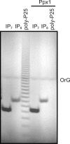
Ppx1 does not hydrolyze inositol pyrophosphates. Recombinant His-Ppx1 (10 ng) was incubated for 30 min at 37 °C with 10 nmol of biochemically synthesized IP7 (5PP-IP5) or IP8 and 100 nmol of poly-P25 that, to the contrary to the inositol pyrophosphate, is fully hydrolyzed. OrG, migration of the Orange G dye.
FIGURE 6.
FIGURE 6.
Comparison of poly-P profiles from ppn1Δ, ppx1Δ, and ddp1Δ mutants. Poly-P was extracted from logarithmic growing cells, and fractions containing 40 μg of total RNA were resolved and visualized with toluidine blue. A, 20% polyacrylamide gel allows the observation of large polymeric species. B, 35% polyacrylamide gel to visualize small poly-P fragments. Knock-out mutants were as follows: ppx1Δ (exopolyphosphatase), ppn1Δ (endopolyphosphatase), and ddp1Δ (diadenosine and diphosphoinositol polyphosphate phosphohydrolase). OrG, migration of the Orange G dye.
FIGURE 7.
FIGURE 7.
DIPP1/2/3 enzymes possess poly-P endophosphatase activity. A, recombinant His-tagged Ddp1, DIPP1, DIPP2, and DIPP3 (100 ng) were resolved on a 4–12% NuPage gel and visualized by Coomassie Blue staining. B, recombinant enzymes (10 ng) were incubated for 60 min at 37 °C with 200 nmol of poly-P25 in a reaction buffered at the indicated pH. C, DIPP1, DIPP2, and DIPP3 (10 ng) were incubated for the indicated time at 37 °C with 200 nmol of poly-P25 in a reaction buffered at pH 9.0. OrG, migration of the Orange G dye.

Similar articles

Cited by

References

    1. Kornberg A., Rao N. N., Ault-Riché D. (1999) Annu. Rev. Biochem. 68, 89–125 - PubMed
    1. Rao N. N., Gómez-García M. R., Kornberg A. (2009) Annu. Rev. Biochem. 78, 605–647 - PubMed
    1. Brown M. R., Kornberg A. (2008) Trends Biochem. Sci. 33, 284–290 - PubMed
    1. Müller F., Mutch N. J., Schenk W. A., Smith S. A., Esterl L., Spronk H. M., Schmidbauer S., Gahl W. A., Morrissey J. H., Renné T. (2009) Cell 139, 1143–1156 - PMC - PubMed
    1. Zhang H., Gómez-García M. R., Shi X., Rao N. N., Kornberg A. (2007) Proc. Natl. Acad. Sci. U.S.A. 104, 16486–16491 - PMC - PubMed

Publication types

MeSH terms

Substances