. 2011 Oct 15;20(20):3943-52.
doi: 10.1093/hmg/ddr313. Epub 2011 Jul 20.

Pitx1 haploinsufficiency causes clubfoot in humans and a clubfoot-like phenotype in mice

Affiliations

Pitx1 haploinsufficiency causes clubfoot in humans and a clubfoot-like phenotype in mice

David M Alvarado et al. Hum Mol Genet. .

Abstract

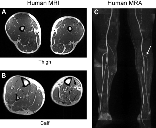
Clubfoot affects 1 in 1000 live births, although little is known about its genetic or developmental basis. We recently identified a missense mutation in the PITX1 bicoid homeodomain transcription factor in a family with a spectrum of lower extremity abnormalities, including clubfoot. Because the E130K mutation reduced PITX1 activity, we hypothesized that PITX1 haploinsufficiency could also cause clubfoot. Using copy number analysis, we identified a 241 kb chromosome 5q31 microdeletion involving PITX1 in a patient with isolated familial clubfoot. The PITX1 deletion segregated with autosomal dominant clubfoot over three generations. To study the role of PITX1 haploinsufficiency in clubfoot pathogenesis, we began to breed Pitx1 knockout mice. Although Pitx1(+/-) mice were previously reported to be normal, clubfoot was observed in 20 of 225 Pitx1(+/-) mice, resulting in an 8.9% penetrance. Clubfoot was unilateral in 16 of the 20 affected Pitx1(+/-) mice, with the right and left limbs equally affected, in contrast to right-sided predominant hindlimb abnormalities previously noted with complete loss of Pitx1. Peroneal artery hypoplasia occurred in the clubfoot limb and corresponded spatially with small lateral muscle compartments. Tibial and fibular bone volumes were also reduced. Skeletal muscle gene expression was significantly reduced in Pitx1(-/-) E12.5 hindlimb buds compared with the wild-type, suggesting that muscle hypoplasia was due to abnormal early muscle development and not disuse atrophy. Our morphological data suggest that PITX1 haploinsufficiency may cause a developmental field defect preferentially affecting the lateral lower leg, a theory that accounts for similar findings in human clubfoot.


Figures

Figure 1.
Figure 1.
Chromosome 5q31 microdeletion containing PITX1 is present in family with isolated clubfoot. (A) Chromosome 5q31 region showing 241 kb deletion at chr5:134222383–134463022 (hg18 build of the UCSC genome browser), involving 124 markers with decreased log2 ratios that was detected in the proband. Four RefSeq genes are located within the interval, including PITX1. (B) The chromosome 5q31 microdeletion segregates with clubfoot in family 5437. Black affection status indicates isolated clubfoot, del indicates deletion and WT indicates normal copy number. (C) Proband from family 5437 with untreated bilateral clubfoot. (D) Three generations of family 5437 with the chromosome 5q31 microdeletion showing surgically treated clubfoot in mother (left), untreated proband (middle) and surgically treated maternal grandmother (right).
Figure 2.
Figure 2.
Muscle volume and vascular abnormalities in the affected clubfoot limb of an adult patient with PITX1 E130K mutation. (A) MRI shows hypoplasia of the left clubfoot limb (shown on the right) compared with the unaffected right leg on transverse sections. (B) Images obtained at calf level show more severe involvement than those taken at the thigh. Muscle hypoplasia is present throughout but is most prominent in the lateral and anterior compartments that also show a corresponding increase in fat tissue (black arrow). (C) Magnetic resonance angiograph of same patient showing reduced perfusion of the anterior tibial (white arrow) and peroneal arteries on the affected left leg compared with the right.
Figure 3.
Figure 3.
Clubfoot-like phenotype is present in some Pitx1 haploinsufficient mice. (A) Lateral view of unaffected hindlimb of Pitx1+/ mouse. (B) Lateral view of affected hindlimb of Pitx1+/ mouse. (C) Dorsal view of unaffected (left) and clubfoot-affected (right) hindlimbs. (D) MicroCT image comparing clubfoot-like right limb of an affected mouse (gray) to unaffected right limb of a control mouse (white) demonstrating forefoot cavus in the frontal plane and (E) hindfoot equinus in the sagittal plane.
Figure 4.
Figure 4.
Peroneal artery hypoplasia is present in the clubfoot-like limb of Pitx1+/− affected mice. Magnetic resonance angiograph of unaffected (A and C) and affected (B and D) Pitx1+/− hindlimbs. The peroneal artery (white arrows) is hypoplastic in affected hindlimbs. There are no apparent anomalies in the anterior tibial or posterior tibial arteries.
Figure 5.
Figure 5.
Tibial volumes are reduced in mouse Pitx1 haploinsufficient clubfoot-like limbs. MicroCT measurements of mouse hindlimb tibia BV indicate (A) decreased average tibia BVs in all Pitx1+/− hindlimbs (unaffected mice and affected clubfoot mice) compared with wild-type controls. These differences correlate with overall reduction in size of Pitx1+/− mice (see text). Average tibia BV of clubfoot-affected limb is smaller than that of the unaffected limb (right side of dotted line). (B) Wild-type and Pitx1+/− unaffected mouse tibia BVs do not differ between left and right within an individual mouse. However, Pitx1+/− unilateral clubfoot-affected mice always have reduced tibia BV in the clubfoot limb compared with the unaffected limbs.
Figure 6.
Figure 6.
Muscle volumes are reduced in mouse Pitx1 haploinsufficient clubfoot-like limbs. (A) Magnetic resonance image of Pitx1+/− unaffected (right) and clubfoot-affected (left) hindlimbs. Tibialis anterior and peroneus muscles (white arrow heads) are hypoplastic in affected hindlimbs while there is a corresponding increase in fat in the lateral compartment (white arrow). (B) Quantitative measurements show that total volume and muscle volume are reduced ∼20% in Pitx1+/− affected hindlimbs compared with the contralateral unaffected limbs. Fat volume is increased in affected hindlimbs compared with the unaffected limbs.

Similar articles

Cited by

References

    1. Wynne-Davies R. Genetic and environmental factors in the etiology of talipes equinovarus. Clin. Orthop. Relat. Res. 1972;84:9–13. - PubMed
    1. Brewer C., Holloway S., Zawalnyski P., Schinzel A., FitzPatrick D. A chromosomal deletion map of human malformations. Am. J. Hum. Genet. 1998;63:1153–1159. - PMC - PubMed
    1. Gurnett C.A., Boehm S., Connolly A., Reimschisel T., Dobbs M.B. Impact of congenital talipes equinovarus etiology on treatment outcomes. Dev. Med. Child Neurol. 2008;50:498–502. - PubMed
    1. Wynne-Davies R. Family studies and the cause of congenital club foot. Talipes equinovarus, talipes calcaneo-valgus and metatarsus varus. J. Bone Joint Surg. Br. 1964;46:445–463. - PubMed
    1. Hootnick D.R., Levinsohn E.M., Crider R.J., Packard D.S., Jr Congenital arterial malformations associated with clubfoot. A report of two cases. Clin. Orthop. Relat. Res. 1982;167:160–163. - PubMed

Publication types

MeSH terms

Substances

Associated data