. 2011 Jul 20;476(7358):101-4.
doi: 10.1038/nature10239.

DMRT1 prevents female reprogramming in the postnatal mammalian testis

Affiliations

DMRT1 prevents female reprogramming in the postnatal mammalian testis

Clinton K Matson et al. Nature. .

Erratum in

  • Nature. 2011 Sep 8;477(7363):238

Abstract

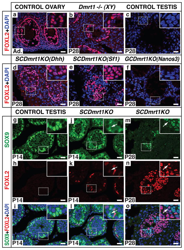
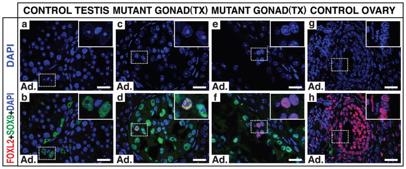
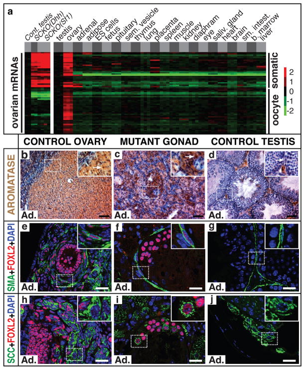
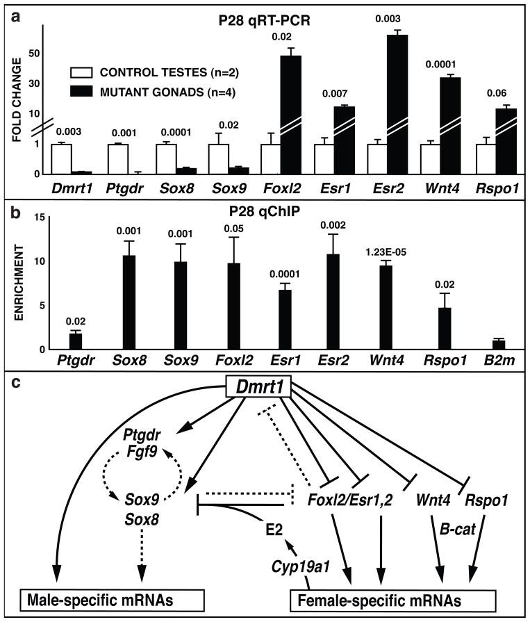
Sex in mammals is determined in the fetal gonad by the presence or absence of the Y chromosome gene Sry, which controls whether bipotential precursor cells differentiate into testicular Sertoli cells or ovarian granulosa cells. This pivotal decision in a single gonadal cell type ultimately controls sexual differentiation throughout the body. Sex determination can be viewed as a battle for primacy in the fetal gonad between a male regulatory gene network in which Sry activates Sox9 and a female network involving WNT/β-catenin signalling. In females the primary sex-determining decision is not final: loss of the FOXL2 transcription factor in adult granulosa cells can reprogram granulosa cells into Sertoli cells. Here we show that sexual fate is also surprisingly labile in the testis: loss of the DMRT1 transcription factor in mouse Sertoli cells, even in adults, activates Foxl2 and reprograms Sertoli cells into granulosa cells. In this environment, theca cells form, oestrogen is produced and germ cells appear feminized. Thus Dmrt1 is essential to maintain mammalian testis determination, and competing regulatory networks maintain gonadal sex long after the fetal choice between male and female. Dmrt1 and Foxl2 are conserved throughout vertebrates and Dmrt1-related sexual regulators are conserved throughout metazoans. Antagonism between Dmrt1 and Foxl2 for control of gonadal sex may therefore extend beyond mammals. Reprogramming due to loss of Dmrt1 also may help explain the aetiology of human syndromes linked to DMRT1, including disorders of sexual differentiation and testicular cancer.


Figures

Figure 1
Figure 1. DMRT1 maintains SOX9 and suppresses FOXL2 expression in postnatal Sertoli cells
(a–c) FOXL2 expression detected by immunofluorescence (IF) in adult granulosa and theca cells of control ovary (a) and intratubular cells of Dmrt1 null testis at postnatal day 28 (P28) (b) but not in control testis (c). (d–f) FOXL2 is robustly expressed when Dmrt1 is mutated in foetal Sertoli cells with Dhh-cre (d) or Sf1-cre (e) but not when Dmrt1 is mutated in foetal germ cells with Nanos3-cre (f). (g–o) Timing of FOXL2 expression. FOXL2 is absent from control testis at P14 (g–i). Cells expressing FOXL2 or FOXL2 and SOX9 (arrowheads) are present in SCDmrt1KO testis at P14 (j–l). FOXL2-positive cells are abundant in SCDmrt1KO testis at P28 and most cells no longer express SOX9 (m–o). Scale bars: 20 mm.
Figure 2
Figure 2. Sertoli-to-granulosa transdifferentiation in the adult testis
Expression of FOXL2 and SOX9 one month after tamoxifen injection into Dmrt1flox/flox adult males (8 weeks and older) carrying inducible ubiquitous cre transgene UBC-cre/ERT2. (a,b) Sertoli cells in control testis express SOX9 but not FOXL2. (c–f) Mutant testis has Sertoli-like cells expressing SOX9 or SOX9 and FOXL2 (inset, d) and granulosa-like cells expressing only FOXL2 (inset, f). (g,h) FOXL2-positive cells in control ovary have DAPI morphology similar to FOXL2 single-positive cells of mutant testis. FOXL2-positive cells in mutant testis resemble granulosa cells: they lack the tripartite nucleoli of Sertoli cells, have smaller and more rounded nuclei, and have more punctate DAPI staining. UBC-cre/ERT2 also deletes Dmrt1 in germ cells, causing precocious meiosis; after one month germ cells are nearly absent. Scale bars: 20 μm.
Figure 3
Figure 3. Feminization of SCDmrt1KO XY gonads
(a) Expression of ovary-enriched mRNAs with expression profiles similar to Foxl2 (see SI). mRNAs labeled “somatic” were enriched in ovarian somatic cells; those labeled “oocyte” were enriched in female germ cells. See Supplemental Fig. 6 for higher resolution image. (b–d) IHC detection of CYP19A1/Aromatase expression in follicles of control adult ovary (a) and in adult XY SCDmrt1KO gonad (b) but only in interstitial Leydig cells of control testis (c). Arrows indicate Aromatase-positive granulosa cells in ovary and mutant gonad and negative Sertoli cell in control testis. Scale bars: 50 μm. (e–g) IF detection of smooth muscle actin (SMA) and FOXL2. Ovarian theca cells (inset, e) are elongated cells expressing both proteins; similar cells are present in mutant gonads (f); peritubular myoid cells in control testes express SMA and not FOXL2 (g). Scale bars: 20 μm. (h–j) IF detection of cells coexpressing FOXL2 in the nucleus and steroidogenic enzyme CYP11A1/SCC at high levels in the cytoplasm in control ovary (h) and XY Dmrt1KO gonads (i). SCC-positive cells in control testis (j) are interstitial Leydig cells. Mutant gonads were SCDmrt1KO(Dhh). Ad.=adult. Scale bars: 20μm.
Figure 4
Figure 4. DMRT1 regulation of postnatal gene expression
(a) qRT-PCR analysis of sex-determining genes at P28. Significance of expression changes is indicated (Students t-test). Mutant gonads were SCDmrt1KO(Sf1); SCDmrt1KO(Dhh) mutant gonads and equivalent expression changes. (b) qChIP analysis of DMRT1 DNA binding in P28 testes. Significance of enrichment relative to B2m (Students t-test) is shown. (c) Model for regulation by postnatal sex maintenance by DMRT1. Proposed direct regulation based on ChIP and mRNA expression data is indicated by solid lines; indirect or potential regulation is indicated by dashed lines. (Model adapted from Veitia).

Comment in

References

    1. Koopman P, Gubbay J, Vivian N, Goodfellow P, Lovell-Badge R. Male development of chromosomally female mice transgenic for Sry. Nature. 1991;351:117–121. doi: 10.1038/351117a0. - DOI - PubMed
    1. Uhlenhaut NH, et al. Somatic sex reprogramming of adult ovaries to testes by FOXL2 ablation. Cell. 2009;139:1130–1142. doi: 10.1016/j.cell.2009.11.021. S0092-8674(09)01433-0 [pii] - DOI - PubMed
    1. Raymond CS, et al. Evidence for evolutionary conservation of sex-determining genes. Nature. 1998;391:691–695. doi: 10.1038/35618. - DOI - PubMed
    1. Loffler KA, Zarkower D, Koopman P. Etiology of ovarian failure in blepharophimosis ptosis epicanthus inversus syndrome: FOXL2 is a conserved, early-acting gene in vertebrate ovarian development. Endocrinology. 2003;144:3237–3243. - PubMed
    1. Raymond CS, Kettlewell JR, Hirsch B, Bardwell VJ, Zarkower D. Expression of Dmrt1 in the genital ridge of mouse and chicken embryos suggests a role in vertebrate sexual development. Dev Biol. 1999;215:208–220. doi: 10.1006/dbio.1999.9461. S0012-1606(99)99461-7 [pii] - DOI - PubMed

Publication types

MeSH terms

Associated data