. 2011 Jul 20;475(7356):343-7.
doi: 10.1038/nature10244.

Type VI secretion delivers bacteriolytic effectors to target cells

Affiliations

Type VI secretion delivers bacteriolytic effectors to target cells

Alistair B Russell et al. Nature. .

Abstract

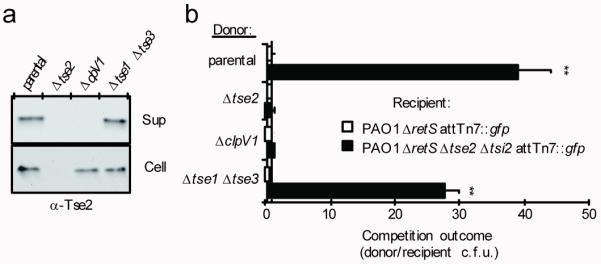
Peptidoglycan is the major structural constituent of the bacterial cell wall, forming a meshwork outside the cytoplasmic membrane that maintains cell shape and prevents lysis. In Gram-negative bacteria, peptidoglycan is located in the periplasm, where it is protected from exogenous lytic enzymes by the outer membrane. Here we show that the type VI secretion system of Pseudomonas aeruginosa breaches this barrier to deliver two effector proteins, Tse1 and Tse3, to the periplasm of recipient cells. In this compartment, the effectors hydrolyse peptidoglycan, thereby providing a fitness advantage for P. aeruginosa cells in competition with other bacteria. To protect itself from lysis by Tse1 and Tse3, P. aeruginosa uses specific periplasmically localized immunity proteins. The requirement for these immunity proteins depends on intercellular self-intoxication through an active type VI secretion system, indicating a mechanism for export whereby effectors do not access donor cell periplasm in transit.


Figures

Figure 1
Figure 1. Tse1 and Tse3 are lytic proteins belonging to amidase and muramidase enzyme families
a. Genomic organization of tse1 and tse3 and their homology with characterized amidase and muramidase enzymes, respectively. Highly conserved (boxed) and catalytic (starred) residues of the respective enzyme families are indicated. SWISS-PROT entry names for the proteins shown are: Tse1 (Q9I2Q1_PSEAE), Spr (SPR_ECOLI), P60 (P60_LISIN), Tse3 (Q9HYC5_PSEAE), GEWL (LYG_ANSAN), Slt70 (SLT_ECOLI). See Supplementary Fig. 1 for full alignments. b,d. Partial HPLC chromatograms of sodium borohydride-reduced soluble E. coli peptidoglycan products resulting from (b) digestion with Tse1 and subsequent cleavage with cellosyl or (d) digestion with Tse3 alone. Peak assignments were made based on MS; predicted structures are shown schematically with hexagons and circles corresponding to sugars and amino acid residues, respectively. Reduced sugar moieties are shown with grey fill. Full chromatograms and MS data are provided in the supplement (Supplementary Fig. 3 and Supplementary Table 1). c. Simplified representation of Gram-negative peptidoglycan showing cleavage sites of Tse1 and Tse3 based on data summarized in b and d. e. Growth in liquid media of E. coli producing the indicated peri-Tse proteins. Periplasmic localization was achieved by fusion to the PelB leader sequence. Cultures were induced at the indicated time (arrow). Error bars ± s.d. (n=3). f. Representative micrographs of strains shown in e acquired prior to complete lysis. The lipophilic dye TMA-DPH is used to highlight the cellular membranes. Supplementary Fig. 5 contains the full microscopic fields from which these images were derived. All images were acquired at the same magnification. Scale bar = 2 μm.
Figure 2
Figure 2. Tse1 and Tse3 are not required for Tse2 export or transfer to recipient cells via the T6S apparatus
a. Western blot analysis of supernatant (Sup) and cell-associated (Cell) fractions of the indicated P. aeruginosa strains. The parental background for all experiments represented in this figure is PAO1 ΔretS, a strain in which the H1-T6SS is activated constitutively,. b. Growth competition assays between the indicated donor and recipient strains under T6S-conducive conditions. Experiments were initiated with equal colony forming units (c.f.u.) of donor and recipient bacteria as denoted by the dashed line. The ΔclpV1 strain is a T6S-deficient control. Asterisks indicate significant differences in competition outcome between recipient strains against the same donor strain. **P < 0.01. Error bars ± s.d. (n=3).
Figure 3
Figure 3. Tsi1 and Tsi3 provide immunity to cognate toxins
a. Western blot analysis of hexahistidine-tagged Tse proteins (–His6) in total and bead-associated fractions of an α-VSV–G (vesicular stomatitis virus glycoprotein) immunoprecipitation of VSV–G epitope fused Tsi proteins (–V) from E. coli. b. Growth of E. coli harboring a vector expressing the indicated tse gene (top panels) or vectors expressing the indicated tse and tsi genes (bottom panels). Numbers at top indicate 10-fold serial dilutions. c. Fluorescence micrographs showing colony growth of the indicated strains. The parental background for this experiment was PAO1 ΔretS attTn7::gfp. Growth of the Δtsi strains was rescued by the addition of 1.0% w/v NaCl to the underlying medium. For quantification of data and complementation analyses see Supplementary Fig. 7. d. Replication rates of the indicated P. aeruginosa strains in liquid medium of low osmolarity formulated as in c. The parental strain used in this experiment was PAO1 ΔretS. Error bars ± s.d. (n=3).
Figure 4
Figure 4. Tse1 and Tse3 delivered to the periplasm provide a fitness advantage to donor cells
a. Western blot analyses of cytoplasmic (Cyto) and periplasmic (Peri) fractions of P. aeruginosa strains producing Tsi1–V, Tsi3–V or Tsi3–SS–V. Equivalent ratios of the Cyto and Peri samples were loaded in each panel. RNA polymerase (RNAP) and β-lactamase (β-lac) enzymes were used as cytoplasmic and periplasmic fractionation controls, respectively. The presence of Tsi3–a predicted outer membrane lipoprotein–in the periplasmic fraction is consistent with previous studies utilizing this method of fractionation. b. Growth competition assays between the indicated donor and recipient strains under T6S-conducive conditions. Experiments were initiated with equal c.f.u. of donor and recipient bacteria as denoted by the dashed line. The parental strain used in this experiment was PAO1 ΔretS. All donor strains were modified at the attB site with lacZ. Asterisks indicate outcomes significantly different than parental versus Δtse3 Δtsi3 (top bar). Error bars ± s.d. (n=4). **P < 0.01. c. Lysis of EDTA-permeabilized or intact P. aeruginosa cells with equal quantities of Tse1, Tse1*, or Lysozyme (Ly). Lysis was normalized to a buffer control. Error bars ± s.d. (n=3). d. Competitive growth of P. aeruginosa against P. putida on solid (open circles) or in liquid (filled circles) medium. Competition outcome was defined as the c.f.u. ratio (P. aeruginosa/P. putida) divided by the initial ratio. The dotted line represents the boundary between competitions that increase in P. aeruginosa relative to P. putida (above the line) and those that increase in P. putida relative to P. aeruginosa (below the line). The parental strain used in this experiment was P. aeruginosa PAO1. Asterisks above competitions denote those where the outcome (P. aeruginosa/P. putida) was significantly less than the parental (P < 0.05). Horizontal bars denote the average value for each dataset (n=5).
Figure 5
Figure 5. Proposed mechanism of T6S-dependent delivery of effector proteins
The schematic depicts the junction between competing bacteria, with a donor cell delivering the Tse effector proteins through the T6S apparatus (grey tube) to recipient cell periplasm. Effector and immunity proteins are shown as circles and rounded rectangles, respectively. Bonds in the peptidoglycan that are predicted targets of the effector proteins are highlighted (red). Cytoplasm (C), inner membrane (IM), periplasm (P), and outer membrane (OM) of both bacteria are shown.

Comment in

References

Online-only methods references

    1. Cardona ST, Valvano MA. An expression vector containing a rhamnose-inducible promoter provides tightly regulated gene expression in Burkholderia cenocepacia. Plasmid. 2005;54:219–228. - PubMed
    1. Hsu F, Schwarz S, Mougous JD. TagR promotes PpkA-catalysed type VI secretion activation in Pseudomonas aeruginosa. Mol Microbiol. 2009;72:1111–1125. - PMC - PubMed
    1. Rietsch A, Vallet-Gely I, Dove SL, Mekalanos JJ. ExsE, a secreted regulator of type III secretion genes in Pseudomonas aeruginosa. Proc Natl Acad Sci U S A. 2005;102:8006–8011. - PMC - PubMed
    1. Horton RM, et al. Gene splicing by overlap extension. Methods in enzymology. 1993;217:270–279. - PubMed
    1. Wood PM. Periplasmic location of the terminal reductase in nitrite respiration. FEBS letters. 1978;92:214–218. doi:0014-5793(78)80757-1 [pii] - PubMed

References

    1. Hayes CS, Aoki SK, Low DA. Bacterial contact-dependent delivery systems. Annual review of genetics. 2010;44:71–90. doi:10.1146/annurev.genet.42.110807.091449. - PubMed
    1. Hibbing ME, Fuqua C, Parsek MR, Peterson SB. Bacterial competition: surviving and thriving in the microbial jungle. Nat Rev Microbiol. 2010;8:15–25. doi:nrmicro2259 [pii] 10.1038/nrmicro2259. - PMC - PubMed
    1. Grundling A, Schneewind O. Cross-linked peptidoglycan mediates lysostaphin binding to the cell wall envelope of Staphylococcus aureus. J Bacteriol. 2006;188:2463–2472. doi:188/7/2463 [pii] 10.1128/JB.188.7.2463-2472.2006. - PMC - PubMed
    1. Vollmer W, Pilsl H, Hantke K, Holtje JV, Braun V. Pesticin displays muramidase activity. J Bacteriol. 1997;179:1580–1583. - PMC - PubMed
    1. Brotz H, Bierbaum G, Markus A, Molitor E, Sahl HG. Mode of action of the lantibiotic mersacidin: inhibition of peptidoglycan biosynthesis via a novel mechanism? Antimicrob Agents Chemother. 1995;39:714–719. - PMC - PubMed

Publication types

MeSH terms