. 2011:2011:405194.
doi: 10.1155/2011/405194. Epub 2011 May 31.

Protective Action of Neurotrophic Factors and Estrogen against Oxidative Stress-Mediated Neurodegeneration

Affiliations

Protective Action of Neurotrophic Factors and Estrogen against Oxidative Stress-Mediated Neurodegeneration

Tadahiro Numakawa et al. J Toxicol. 2011.

Abstract

Oxidative stress is involved in the pathogenesis of neurodegenerative disorders such as Alzheimer's disease, Parkinson's disease, and Huntington's disease. Low levels of reactive oxygen species (ROS) and reactive nitrogen species (RNS) are important for maintenance of neuronal function, though elevated levels lead to neuronal cell death. A complex series of events including excitotoxicity, Ca(2+) overload, and mitochondrial dysfunction contributes to oxidative stress-mediated neurodegeneration. As expected, many antioxidants like phytochemicals and vitamins are known to reduce oxidative toxicity. Additionally, growing evidence indicates that neurotrophic factors such as brain-derived neurotrophic factor (BDNF) and estrogens significantly prevent neuronal damage caused by oxidative stress. Here, we review and discuss recent studies addressing the protective mechanisms of neurotrophic factors and estrogen within this system.


Figures

Figure 1
Figure 1
Mechanisms underlying oxidative stress-mediated neuronal apoptosis. Accumulation of oxidative stress is involved in the development/progression of neurodegenerative diseases. A number of events including excitotoxicity, mitochondrial dysfunction, Ca2+ overload, and endoplasmic reticulum stress are associated with excess reactive oxygen species (ROS) and reactive nitrogen species (RNS) generation. High levels of ROS/RNS lead to oxidation of proteins, lipids, and DNA. Oxidized lipids induce damage of the ubiquitin-proteasome system (UPS). The UPS dysfunction and oxidation of proteins result in aggregation of proteins, recognized as a hallmark of several neurodegenerative diseases. Under oxidative stress, death signaling pathways (p53, mitogen-activated protein kinase (MAPK), etc.) are activated. Activation of p53 leads to induction of proapoptotic proteins such as Bax and p53-upregulated modulator of apoptosis (PUMA), followed by translocation of these proteins into mitochondria. Finally, mitochondrial cytochrome c is released, which then stimulates the activation of caspase 9/caspase 3. Alternatively, mitochondria secrete apoptosis-inducing factor (AIF), leading to caspase-independent apoptosis. As shown, recent studies suggest antioxidant effects of phytochemicals, vitamin E, estrogen, and neurotrophic factors including brain-derived neurotrophic factor (BDNF), glial cell line-derived neurotrophic factor (GDNF), and hepatocyte growth factor (HGF), leading to increased preservation of neuronal function.
Figure 2
Figure 2
17β-estradiol prevents cortical neurons from cell death caused by H2O2 exposure. Dissociated cortical neurons were prepared from cerebral cortex of postnatal 2-day-old rats. At 6 days in vitro, 17β-estradiol was applied at indicated concentrations. Twenty-four hours later, H2O2 (final 50 μM) was added to induce cell death. Following an additional twelve-hour culture, cell survival was determined using an MTT (tetrazolium salt) assay. Data represent mean ± S.D. (n = 6). ***P < .001 versus control (no H2O2). ### P < .001 versus no estradiol + H2O2.
Figure 3
Figure 3
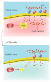
17β-estradiol inhibits neuronal cell death under oxidative stress via reducing the series of events evoked by exposure to H2O2, including overactivation of the ERK signaling and overload of Ca2+. Upper: After H2O2 addition, marked phosphorylated (activated) ERK (pERK) and resultant increase in intracellular Ca2+ concentration were observed, resulting in cell death. Lower: Pretreatment with 17β-estradiol induced downregulation of ionotropic glutamate receptors via decreasing ERK activation, while also serving to decrease levels of Ca2+ influx triggered by H2O2. Such a decrease in glutamate receptor expression and intracellular Ca2+ was also confirmed in the presence of U0126, an inhibitor of ERK signaling. As expected, chronic 17β-estradiol reduced levels of pERK stimulated by H2O2. A blockade of glutamate receptors rescued cortical cells from H2O2-dependent death. Therefore, it is possible that 17β-estradiol promotes survival via suppressing glutamate receptor-mediated Ca2+ influx, due to downregulation of ionotropic glutamate receptors [120].

Similar articles

Cited by

References

    1. Sakurada O, Kennedy C, Jehle J, Brown JD, Carbin GL, Sokoloff L. Measurement of local cerebral blood flow with iodo [14C] antipyrine. The American Journal of Physiology. 1978;234(1):H59–H66. - PubMed
    1. Cremer JE, Seville MP. Regional brain blood flow, blood volume, and haematocrit values in the adult rat. Journal of Cerebral Blood Flow and Metabolism. 1983;3(2):254–256. - PubMed
    1. Scremin OU, Sonnenschein RR, Rubinstein EH. Cholinergic cerebral vasodilatation: lack of involvement of cranial parasympathetic nerves. Journal of Cerebral Blood Flow and Metabolism. 1983;3(3):362–368. - PubMed
    1. Sokoloff L, Reivich M, Kennedy C, et al. The [C]deoxyglucose method for the measurement of local cerebral glucose utilization: theory, procedure, and normal values in the conscious and anesthetized albino rat. Journal of Neurochemistry. 1977;28(5):897–916. - PubMed
    1. Sokoloff L. Localization of functional activity in the central nervous system by measurement of glucose utilization with radioactive deoxyglucose. Journal of Cerebral Blood Flow and Metabolism. 1981;1(1):7–36. - PubMed