Cytotoxicity of environmentally relevant concentrations of aluminum in murine thymocytes and lymphocytes
- PMID: 21776265
- PMCID: PMC3135276
- DOI: 10.1155/2011/796719
Cytotoxicity of environmentally relevant concentrations of aluminum in murine thymocytes and lymphocytes
Abstract
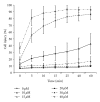
The effects of low concentrations of aluminum chloride on thymocytes and lymphocytes acutely dissociated from young mice were studied using flow cytometry with a DNA-binding dye. We demonstrate a rapid and dose-dependent injury in murine thymocytes and lymphocytes resulting from exposure to aluminum, as indicated by an increase in the entry into the cell of the DNA-binding dye, propidium iodine. A 60-minute exposure to 10 μM AlCl(3) caused damage of about 5% of thymocytes, while 50% were injured after 10 minutes at 20 μM. Nearly all thymocytes showed evidence of damage at 30 μM AlCl(3) after only 5 minutes of incubation. In lymphocytes, injury was observed at 15 μM AlCl(3) and less than 50% of cells were injured after a 60-minute exposure to 20 μM. Injury only rarely proceeded to rapid cell death and was associated with cell swelling. These results suggest that aluminum has cytotoxic effects on cells of the immune system.
Figures


Similar articles
-
A natural immunity-activating plant lectin, Viscum album agglutinin-I, induces apoptosis in human lymphocytes, monocytes, monocytic THP-1 cells and murine thymocytes.Nat Immun. 1996-1997;15(6):295-311. Nat Immun. 1996. PMID: 9523281
-
Deltamethrin induced an apoptogenic signalling pathway in murine thymocytes : exploring the molecular mechanism.J Appl Toxicol. 2014 Dec;34(12):1303-10. doi: 10.1002/jat.2948. Epub 2013 Nov 12. J Appl Toxicol. 2014. PMID: 24217896
-
In vitro apoptosis of lymphocytes after exposure to levels of corticosterone observed following submaximal exercise.J Sports Med Phys Fitness. 1999 Dec;39(4):269-74. J Sports Med Phys Fitness. 1999. PMID: 10726425
-
Total allowable concentrations of monomeric inorganic aluminum and hydrated aluminum silicates in drinking water.Crit Rev Toxicol. 2012 May;42(5):358-442. doi: 10.3109/10408444.2012.674101. Crit Rev Toxicol. 2012. PMID: 22512666 Review.
-
Dose-dependent opposite effect of zinc on apoptosis in mouse thymocytes.Int J Immunopharmacol. 1995 Sep;17(9):735-44. doi: 10.1016/0192-0561(95)00063-8. Int J Immunopharmacol. 1995. PMID: 8582785
Cited by
-
Acute Toxic and Genotoxic Effects of Aluminum and Manganese Using In Vitro Models.Toxics. 2021 Jun 30;9(7):153. doi: 10.3390/toxics9070153. Toxics. 2021. PMID: 34208861 Free PMC article.
-
Systematic review of potential health risks posed by pharmaceutical, occupational and consumer exposures to metallic and nanoscale aluminum, aluminum oxides, aluminum hydroxide and its soluble salts.Crit Rev Toxicol. 2014 Oct;44 Suppl 4(Suppl 4):1-80. doi: 10.3109/10408444.2014.934439. Crit Rev Toxicol. 2014. PMID: 25233067 Free PMC article.
-
The Effect of Aluminum Exposure on Maternal Health and Fetal Growth in Rats.Cureus. 2022 Nov 22;14(11):e31775. doi: 10.7759/cureus.31775. eCollection 2022 Nov. Cureus. 2022. PMID: 36425052 Free PMC article.
-
Water-soluble optical sensors: keys to detect aluminium in biological environment.RSC Adv. 2022 May 10;12(22):13950-13970. doi: 10.1039/d2ra01222g. eCollection 2022 May 5. RSC Adv. 2022. PMID: 35558844 Free PMC article. Review.
-
Aluminium toxicosis: a review of toxic actions and effects.Interdiscip Toxicol. 2019 Oct;12(2):45-70. doi: 10.2478/intox-2019-0007. Epub 2020 Feb 20. Interdiscip Toxicol. 2019. PMID: 32206026 Free PMC article. Review.
References
-
- Greger JL, Sutherland JE. Aluminum exposure and metabolism. Critical Reviews in Clinical Laboratory Sciences. 1997;34(5):439–474. - PubMed
-
- Haug AE. Molecular aspects of aluminum toxicity. Critical Reviews in Plant Sciences. 1984;1:345–373.
-
- Segev G, Bandt C, Francey T, Cowgill LD. Aluminum toxicity following administration of aluminum-based phosphate binders in 2 dogs with renal failure. Journal of Veterinary Internal Medicine. 2008;22(6):1432–1435. - PubMed
-
- Heaf JG. Bone disease after renal transplantation. Transplantation. 2003;75(3):315–325. - PubMed
-
- Bishop NJ, Morley R, Philip Day J, Lucas A. Aluminum neurotoxicity in preterm infants receiving intravenous-feeding solutions. New England Journal of Medicine. 1997;336(22):1557–1561. - PubMed
LinkOut - more resources
Full Text Sources

