

Radiation protection in pediatric radiology
- PMID: 21776310
- PMCID: PMC3132617
- DOI: 10.3238/arztebl.2011.0407
Radiation protection in pediatric radiology
Abstract
Background: The German Federal Law on Radiation Control contains no special provisions for X-ray studies in children and adolescents, even though exposure to ionizing radiation must be kept especially low in young persons, because their tissues are highly radiosensitive. Children, who have many years left to live, are more likely than adults to develop radiation-induced cancer; also, as future parents, they are at risk for passing on radiation-induced genetic defects to the next generation. Whenever possible, radiological studies on children and adolescents should be of a type that does not involve ionizing radiation, such as ultrasonography or magnetic resonance imaging. Pediatric conventional X-rays and computerized tomography (CT) require special examining techniques and protocols that are adapted to the patient's age and to the indication for the study.
Methods: We selectively review the literature on pediatric dose reduction and discuss our own investigations on the subject as well.
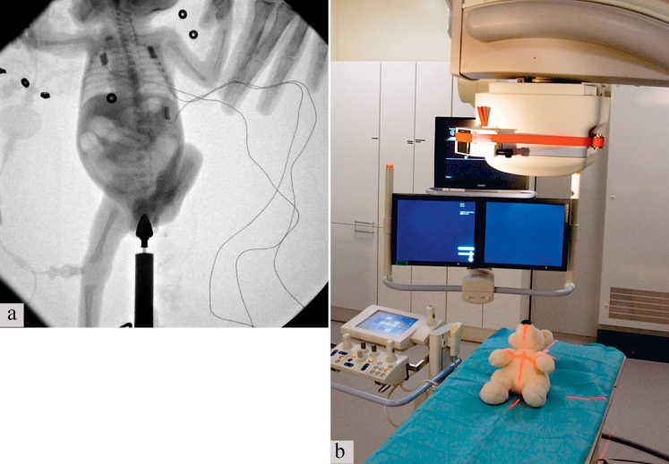
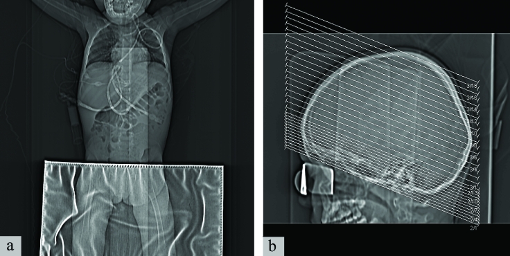
Results: The essential technical prerequisites for lowering the dose of ionizing radiation in conventional X-ray studies include the proper setting of tube voltage, the use of tube filters, suitable patient positioning and fixation, variable use of a scattered-radiation grid, and a modern storage-plate system. In CT studies, the use of age- and indication-adapted protocols can lower radiation exposure by as much as 95%.
Conclusion: There are now many ways to lower the exposure of children and adolescents to ionizing radiation without sacrificing diagnostic reliability. The main factors in lowering exposure are proper attention to clinical indications, the use of special X-ray protocols, the use of alternative imaging studies without ionizing radiation wherever possible, and the expertise of the examiner.
Figures


References
-
- Berington Gonzalez A de, Darby S. Risk of cancer from diagnostic X-rays: estimates for the UK and 14 other countries. Lancet. 2004;363:345–351. - PubMed
-
- Brenner DJ. Estimating cancer risks from pediatric CT: going from qualitative to the quantitative. Pediatr Radiol. 2002;32:228–231. - PubMed
-
- Brenner DJ, Elliston CD. Estimated radiation risks potentially associated with full-body CT screening. Radiology. 2004;232:735–738. - PubMed
-
- Hall EJ. Lessons we have learned from our children: cancer risks from diagnostic radiology. Pediatr Radiol. 2002;32(10):700–706. - PubMed
-
- Kuttesch JF, Jr, Wexler LH, Marcus RB, et al. Second malignancies after Ewing’s sarcoma: radiation dose-dependency of secondary sarcomas. J Clin Oncol. 1996;14(10):2818–2825. - PubMed