. 2011 Sep;118(6):979-87.
doi: 10.1111/j.1471-4159.2011.07392.x. Epub 2011 Aug 12.

Expression of the 2',3'-cAMP-adenosine pathway in astrocytes and microglia

Affiliations

Expression of the 2',3'-cAMP-adenosine pathway in astrocytes and microglia

Jonathan D Verrier et al. J Neurochem. 2011 Sep.

Abstract

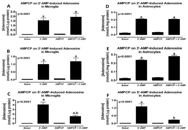
Many organs express the extracellular 3',5'-cAMP-adenosine pathway (conversion of extracellular 3',5'-cAMP to 5'-AMP and 5'-AMP to adenosine). Some organs release 2',3'-cAMP (isomer of 3',5'-cAMP) and convert extracellular 2',3'-cAMP to 2'- and 3'-AMP and convert these AMPs to adenosine (extracellular 2',3'-cAMP-adenosine pathway). As astrocytes and microglia are important participants in the response to brain injury and adenosine is an endogenous neuroprotectant, we investigated whether these extracellular cAMP-adenosine pathways exist in these cell types. 2',3'-, 3',5'-cAMP, 5'-, 3'-, and 2'-AMP were incubated with mouse primary astrocytes or primary microglia for 1 h and purine metabolites were measured in the medium by mass spectrometry. There was little evidence of a 3',5'-cAMP-adenosine pathway in either astrocytes or microglia. In contrast, both cell types converted 2',3'-cAMP to 2'- and 3'-AMP (with 2'-AMP being the predominant product). Although both cell types converted 2'- and 3'-AMP to adenosine, microglia were five- and sevenfold, respectively, more efficient than astrocytes in this regard. Inhibitor studies indicated that the conversion of 2',3'-cAMP to 2'-AMP was mediated by a different ecto-enzyme than that involved in the metabolism of 2',3'-cAMP to 3'-AMP and that although CD73 mediates the conversion of 5'-AMP to adenosine, an alternative ecto-enzyme metabolizes 2'- or 3'-AMP to adenosine.


Conflict of interest statement

No author had a conflict of interest.

Figures

Figure 1
Figure 1
Cell-type characterization of primary microglial cell cultures. Microglial cells were identified using anti-Iba-1 primary and FITC-conjugated secondary antibodies (Green, DAPI, blue; Scale bar = 50 microns) (A), and astrocytes were identified using anti-GFAP primary and RED-conjugated secondary antibodies (Red, DAPI, blue) (B). Also presented is a merged image of the same field of view showing both cell types (C). Quantification of random fields of view reveals that the primary cultures are over 98% Iba-1+ microglial cells with only few residual astrocytes present (***, p<0.001) (D).
Figure 2
Figure 2
Line graphs show the concentration-dependent effects in microglia (A, B, and C) and astrocytes (D, E and F) of 3′,5′-cAMP and 2′,3′-cAMP on extracellular levels of 2′-AMP (A and D), 3′-AMP (B and E) and 5′-AMP (C and F). P-values in panels are from analysis of variance. ap<0.05, compared with basal (0). Values represent means ± SEM for 6 experiments.
Figure 3
Figure 3
Bar graphs illustrate the effects of 3-isobutyl-1-methylxanthine (IBMX, 1 mmol/L; broad spectrum phosphodiesterase inhibitor) and 1,3-dipropyl-8-p-sulfophenylxanthine (DPSPX, 1 mmol/L; ecto-phosphodiesterase inhibitor) in microglia (A and B) and astrocytes (C and D) on the metabolism of 2′,3′-cAMP to 2′-AMP (A and C) and 3′-AMP (B and D). P-values in panels are from analysis of variance. ap<0.05, compared with basal; bp<0.05, compared with no inhibitor. Values represent means ± SEM for 6 experiments.
Figure 4
Figure 4
Line graphs show the concentration-dependent effects in microglia (A, B and C) and astrocytes (D, E and F) of 2′-AMP (A and D), 3′-AMP (B and E) and 5′-AMP (C and F) on extracellular levels of adenosine. P-values in panels are from analysis of variance. ap<0.05, compared with basal (0). Values represent means ± SEM for 6 experiments.
Figure 5
Figure 5
Bar graphs depict the effects in microglia (A, B and C) and astrocytes (D, E and F) of α,β-methylene-adenosine-5′-diphosphate (AMPCP, 0.1 mmol/L; ecto-5′-nucleotidase inhibitor) on metabolism of 2′-AMP (A and D), 3′-AMP (B and E) and 5′-AMP (C and F) to adenosine. P-values in panels are from analysis of variance. ap<0.05, compared with basal (0); bp<0.05, compared with 5′-AMP alone. Values represent means ± SEM for 6 experiments.
Figure 6
Figure 6
Line graphs show the concentration-dependent effects of 3′,5′-cAMP and 2′,3′-cAMP on extracellular levels of adenosine in microglia (A) and astrocytes (B). P-values in panels are from analysis of variance. ap<0.05, compared with basal (0). Values represent means ± SEM for 6 experiments.

References

    1. Azarashvili T, Krestinina O, Galvita A, Grachev D, Baburina Y, Stricker R, Evtodienko Y, Reiser G. Ca2+-dependent permeability transition regulation in rat brain mitochondria by 2′,3′-cyclic nucleotides and 2′,3′-cyclic nucleotide 3′-phosphodiesterase. Am J Physiol Cell Physiol. 2009;296:1428–1439. - PubMed
    1. Beavo JA, Reifsnyder DH. Primary sequence of cyclic nucleotide phosphodiesterase isozymes and the design of selective inhibitors. Trends Pharmacol Sci. 1990;11:150–155. - PubMed
    1. Brundege JM, Diao L, Proctor WR, Dunwiddie TV. The role of cyclic AMP as a precursor of extracellular adenosine in the rat hippocampus. Neuropharmacology. 1997;36:1201–1210. - PubMed
    1. Chiavegatti T, Costa VL, Jr, Araujo MS, Godinho RO. Skeletal muscle expresses the extracellular cyclic AMP-adenosine pathway. Br J Pharmacol. 2008;153:1331–1340. - PMC - PubMed
    1. Chouker A, Thiel M, Lukashev D, Ward JM, Kaufmann I, Apasov S, Sitkovsky MV, Ohta A. Critical role of hypoxia and A2A adenosine receptors in liver tissue-protecting physiological anti-inflammatory pathway. Molecular Medicine. 2008;14:116–123. - PMC - PubMed

Publication types

MeSH terms