. 2011 Jul 21;30(1):69.
doi: 10.1186/1756-9966-30-69.

Application of Benchtop-magnetic resonance imaging in a nude mouse tumor model

Affiliations

Application of Benchtop-magnetic resonance imaging in a nude mouse tumor model

Henrike Caysa et al. J Exp Clin Cancer Res. .

Abstract

Background: MRI plays a key role in the preclinical development of new drugs, diagnostics and their delivery systems. However, very high installation and running costs of existing superconducting MRI machines limit the spread of MRI. The new method of Benchtop-MRI (BT-MRI) has the potential to overcome this limitation due to much lower installation and almost no running costs. However, due to the low field strength and decreased magnet homogeneity it is questionable, whether BT-MRI can achieve sufficient image quality to provide useful information for preclinical in vivo studies. It was the aim of the current study to explore the potential of BT-MRI on tumor models in mice.

Methods: We used a prototype of an in vivo BT-MRI apparatus to visualise organs and tumors and to analyse tumor progression in nude mouse xenograft models of human testicular germ cell tumor and colon carcinoma.

Results: Subcutaneous xenografts were easily identified as relative hypointense areas in transaxial slices of NMR images. Monitoring of tumor progression evaluated by pixel extension analyses based on NMR images correlated with increasing tumor volume calculated by calliper measurement. Gd-BOPTA contrast agent injection resulted in a better differentiation between parts of the urinary tissues and organs due to fast elimination of the agent via kidneys. In addition, interior structuring of tumors could be observed. A strong contrast enhancement within a tumor was associated with a central necrotic/fibrotic area.

Conclusions: BT-MRI provides satisfactory image quality to visualize organs and tumors and to monitor tumor progression and structure in mouse models.


Figures

Figure 1
Figure 1
Prototype of the Benchtop-MRI system "MARAN DRX2" (Oxford Instruments).
Figure 2
Figure 2
Transaxial NMR images of mice (face-down position) bearing two s.c. xenografts; left: 1411HP germ cell tumor, right: DLD-1 colon carcinoma. Images were taken without Gd-BOPTA and 10 min, 20 min and 30 min after i.v. application of Gd-BOPTA. (A): The illustration of renal pelvis was clearly enhanced directly after contrast agent injection in light grey compared to a black central area without Gd-BOPTA. The fast nephritic elimination caused a signal decrease (darker grey) already after 30 min. White arrows point at kidneys. (B): High contrast enhancement in the urinary bladder (white arrow) was identifiable as hypertense area compared to a hypotense one without contrast agent. (C): Subcutaneous xenograft tumors are visible as relative hypointense area (white arrows).
Figure 3
Figure 3
Analysis of contrast agent induced interior structuring of tumours. (A): Transaxial NMR images of a mouse (face-down position) bearing two s.c. xenografts; left: HT29 colon carcinoma, right HCT8 colon carcinoma. Images were taken to the indicated time points after i.v. application of higher dosed Gd-BOPTA (0.1 mmol/kg). A time dependent alteration of contrast enhancement with initial enhancement of the tumor rim followed by a centripetal progression of the signal is observed in the HT29 tumor. The HCT8 tumor was too small for detailed analyses although a time dependent alteration of the signal could also be observed. (upper panel - grayscale, lower panel - pseudocolor) (B): Transaxial NMR images of a mouse (face-down position) bearing two s.c. HT29 xenografts 15 min and 30 min after i.v. application of Gd-BOPTA. One tumor showed strong contrast enhancement and an interior structuring could be observed (white arrow). (C): HE staining of the well structured left HT29 xenograft shown in (A). Depicted is a section at the side of the tumor to represent the whole structure composed of a large central necrotic/fibrotic area (white star) surrounded by peripherally arranged vital tumor cells (white arrow).
Figure 4
Figure 4
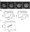
Monitoring of xenograft tumor growth. (A): Transaxial NMR images of a mouse (face-down position) bearing two s.c. xenografts (left: 1411HP germ cell tumor, right: DLD-1 colon carcinoma) analysed over 5 weeks (d13, d20, d27, d34 post cell injection). Depicted images were taken 10 min after i.v. application of Gd-BOPTA. White arrows point at tumors. (B): Following tumor growth of example shown in Figure 4A as analysed by calliper measurements and volume calculation compared to analyses by pixel extension of tumor sections based on NMR images (with or without Gd-BOPTA (CA)). Both tumor volume (V) and tumor section extent (A) comparably increased over the observation period. (C): Correlation of both methods: calculation of tumor growth by calliper measurement (V) and pixel extension analyses based on NMR images (A) of all 12 tumors.

References

    1. Malaterre V, Metz H, Ogorka J, Gurny R, Loggia N, Mader K. Benchtop-magnetic resonance imaging (BT-MRI) characterization of push-pull osmotic controlled release systems. J Control Release. 2009;133:31–36. doi: 10.1016/j.jconrel.2008.09.007. - DOI - PubMed
    1. Metz H, Mader K. Benchtop-NMR and MRI - a new analytical tool in drug delivery research. Int J Pharm. 2008;364:170–175. doi: 10.1016/j.ijpharm.2008.09.033. - DOI - PubMed
    1. Strubing S, Abboud T, Contri RV, Metz H, Mader K. New insights on poly(vinyl acetate)-based coated floating tablets: characterisation of hydration and CO2 generation by benchtop MRI and its relation to drug release and floating strength. Eur J Pharm Biopharm. 2008;69:708–717. doi: 10.1016/j.ejpb.2007.12.009. - DOI - PubMed
    1. Strubing S, Metz H, Mader K. Characterization of poly(vinyl acetate) based floating matrix tablets. J Control Release. 2008;126:149–155. doi: 10.1016/j.jconrel.2007.11.013. - DOI - PubMed
    1. Nitzsche H, Metz H, Lochmann A, Bernstein A, Hause G, Groth T, Mader K. Characterization of scaffolds for tissue engineering by benchtop-magnetic resonance imaging. Tissue Eng Part C Methods. 2009;15:513–521. doi: 10.1089/ten.tec.2008.0488. - DOI - PubMed

Publication types

MeSH terms