. 2011 Nov;104(3):395-403.
doi: 10.1016/j.ymgme.2011.06.002. Epub 2011 Jun 12.

Inverse zonation of hepatocyte transduction with AAV vectors between mice and non-human primates

Affiliations

Inverse zonation of hepatocyte transduction with AAV vectors between mice and non-human primates

Peter Bell et al. Mol Genet Metab. 2011 Nov.

Abstract

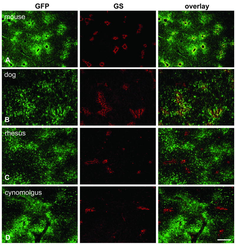
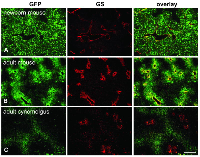
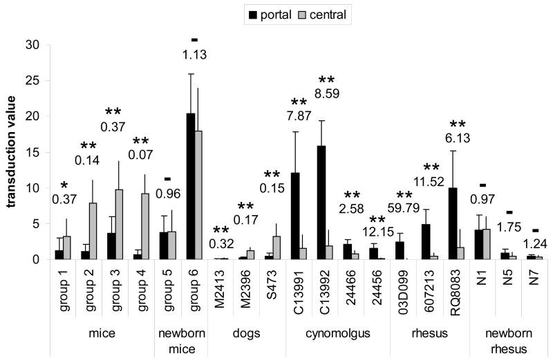
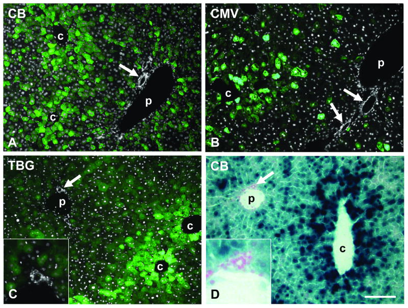
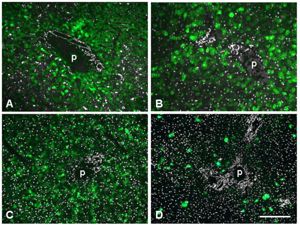
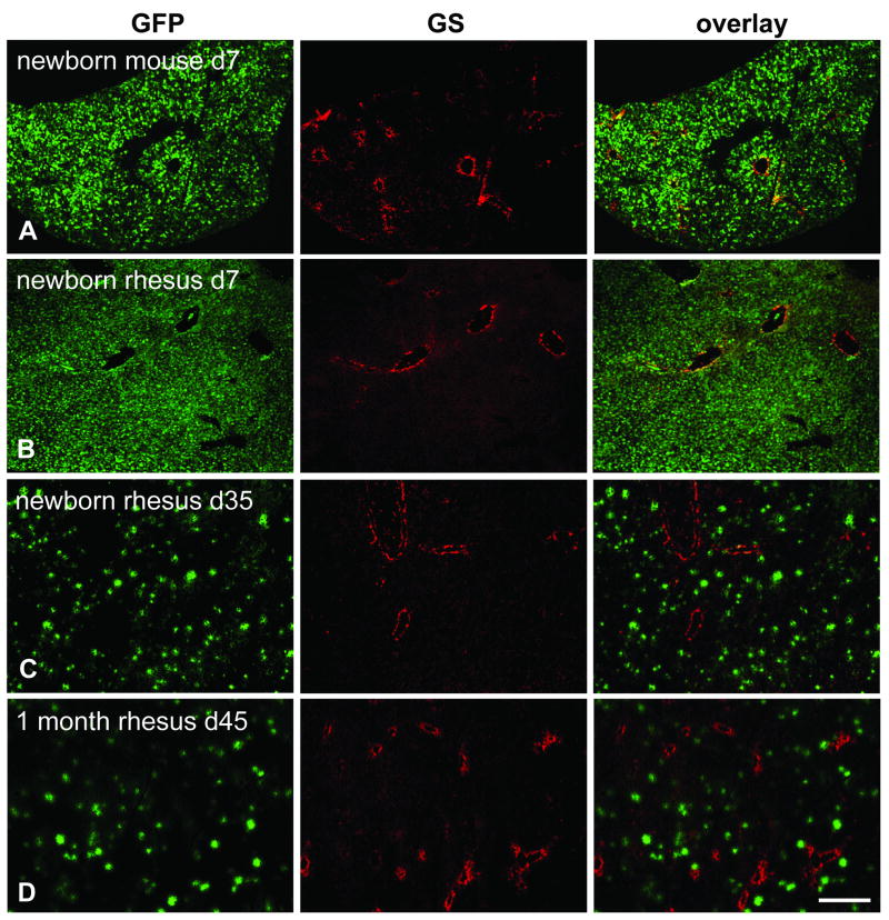
Gene transfer vectors based on adeno-associated virus 8 (AAV8) are highly efficient in liver transduction and can be easily administered by intravenous injection. In mice, AAV8 transduces predominantly hepatocytes near central veins and yields lower transduction levels in hepatocytes in periportal regions. This transduction bias has important implications for gene therapy that aims to correct metabolic liver enzymes because metabolic zonation along the porto-central axis requires the expression of therapeutic proteins within the zone where they are normally localized. In the present study we compared the expression pattern of AAV8 expressing green fluorescent protein (GFP) in liver between mice, dogs, and non-human primates. We confirmed the pericentral dominance in transgene expression in mice with AAV8 when the liver-specific thyroid hormone-binding globulin (TBG) promoter was used but also observed the same expression pattern with the ubiquitous chicken β-actin (CB) and cytomegalovirus (CMV) promoters, suggesting that transduction zonation is not caused by promoter specificity. Predominantly pericentral expression was also found in dogs injected with AAV8. In contrast, in cynomolgus and rhesus macaques the expression pattern from AAV vectors was reversed, i.e. transgene expression was most intense around portal areas and less intense or absent around central veins. Infant rhesus macaques as well as newborn mice injected with AAV8 however showed a random distribution of transgene expression with neither portal nor central transduction bias. Based on the data in monkeys, adult humans treated with AAV vectors are predicted to also express transgenes predominantly in periportal regions whereas infants are likely to show a uniform transduction pattern in liver.


Figures

Fig. 1
Fig. 1
Immunofluorescence staining on liver after gene transfer with AAV8 expressing GFP from the TBG promoter. Sections were stained with antibodies against GFP (green, left column) and glutamine synthetase (GS) as marker for central veins (red, middle column). The overlay of both stains is shown in the right column. All animals were analyzed seven days after vector treatment and received a dose of 3×10¹² GC/kg except for the mouse (1×10¹¹ GC per animal) and the dog (1.82×10¹³ GC/kg). A. Mouse liver (group 2). B. Dog liver (animal S473). C. Juvenile rhesus macaque (animal RQ8082). D. Adult cynomolgus macaque (animal C13992). Scale bar: 400 μm.
Fig. 2
Fig. 2
Immunofluorescence staining for GFP and GS on livers from animals that received self-complementary (sc) vectors expressing GFP. A. Newborn mouse (group 6) injected with 5×10¹⁰ GC of scAAV8.TBG.EGFP and analyzed seven days later. B. Adult mouse (group 4) treated with 3×10⁹ GC of the same vector seven days after injection. C. Adult cynomolgus macaque (24456) injected with 2×10¹² GC/kg of scAAV7 expressing GFP from a CB promoter. Scale bar: 400 μm.
Fig. 3
Fig. 3
Morphometric analysis comparing portal and central transduction levels in mice, dogs, and non-human primates after treatment with vector as adults or newborns. Portal-central ratios of the transduction values are shown above each group or animal. Statistical significance between portal and central transduction values as determined by student t-test is indicated by asterisks (* p = 0.012, ** p < 0.001), no significance by – (p > 0.1). See Table 1 for type of vector, dose, time point and other details. All animals received AAV8 expressing GFP from the TBG promoter with the exception of mice from group 1 (scAAV8 expressing hOTC detected by immunofluorescence staining) and cynomolgus macaques 24466 and 24456 (scAAV7 expressing GFP from CB promoter). Self-complementary vectors have been used in mice groups 1 and 4 and for macaques 24466 and 24456. The transduction value was determined by measuring the average transduced area within a 225 μm radius around either central or portal veins and is reported as percentage per total image area (see Materials and Methods for details). Error bars represent standard deviation.
Fig. 4
Fig. 4
Predominantly pericentral transduction in mouse liver after tail vein injection of AAV8 vectors with different promoters and transgenes at a dose of 1×10¹¹ GC per animal. A – C shows GFP fluorescence (green) overlaid with DAPI staining (white) to demonstrate bile duct and hepatic artery, D shows X-gal staining for β-galactosidase expression. A. GFP expressed from CB promoter (day 14 post injection). B. GFP expressed from CMV promoter (day 9). C. GFP expressed from TBG promoter (day 7). D. nLacZ expressed from CB promoter (day 14). Arrow marks bile duct or hepatic artery (shown enlarged in insets). c and p indicate central and portal vein, respectively. Scale bar: 150 μm.
Fig. 5
Fig. 5
GFP expression in portal areas of liver from non-human primates after administration of 3×10¹² GC/kg of AAV8 expressing GFP from the TBG promoter. Shown is GFP fluorescence (green) overlaid with DAPI staining (white) to demonstrate bile duct and hepatic artery. A. Adult cynomolgus macaque C13991 seven days after injection. B. Adult rhesus macaque 607213 seven days after injection. C. Infant rhesus macaque N1, injected 1 week after birth and analyzed seven days later. D. Infant rhesus macaque N5, injected 1 week after birth and analyzed 35 days later. p indicates portal vein. Scale bar: 200 μm (A, C, D) and 130 μm (B).
Fig. 6
Fig. 6
Immunofluorescence staining for GFP and GS on livers of neonatal mice and infant rhesus macaques. The mouse received 2.5×10¹⁰ GC total and the rhesus macaques 3×10¹² GC/kg of AAV8.TBG.EGFP. A. Mouse (group 5) injected within 24 h after birth and analyzed seven days after treatment. B. Macaque N1 injected 1 week after birth and analyzed seven days later. C. Macaque N5 injected 1 week after birth and analyzed 35 days later. D. Macaque N7 injected 1 month after birth and analyzed 45 days later. Scale bar: 400 μm.

References

    1. Wang L, Wang H, Bell P, McCarter RJ, He J, Calcedo R, Vandenberghe LH, Morizono H, Batshaw ML, Wilson JM. Systematic evaluation of AAV vectors for liver directed gene transfer in murine models. Mol Ther. 2010;18:118–125. - PMC - PubMed
    1. Wang L, Calcedo R, Wang H, Bell P, Grant R, Vandenberghe LH, Sanmiguel J, Morizono H, Batshaw ML, Wilson JM. The pleiotropic effects of natural AAV infections on liver-directed gene transfer in macaques. Mol Ther. 2010;18:126–134. - PMC - PubMed
    1. Gao G, Lu Y, Calcedo R, Grant RL, Bell P, Wang L, Figueredo J, Lock M, Wilson JM. Biology of AAV serotype vectors in liver-directed gene transfer to nonhuman primates. Mol Ther. 2006;13:77–87. - PubMed
    1. Katz NR. Metabolic heterogeneity of hepatocytes across the liver acinus. J Nutr. 1992;122:843–849. - PubMed
    1. Gebhardt R. Metabolic zonation of the liver: regulation and implications for liver function. Pharmacol Ther. 1992;53:275–354. - PubMed

Publication types

Substances