. 2011 Sep 8;118(10):2906-17.
doi: 10.1182/blood-2011-01-331694. Epub 2011 Jul 21.

Pericytes promote endothelial cell survival through induction of autocrine VEGF-A signaling and Bcl-w expression

Affiliations

Pericytes promote endothelial cell survival through induction of autocrine VEGF-A signaling and Bcl-w expression

Marcela Franco et al. Blood. .

Abstract

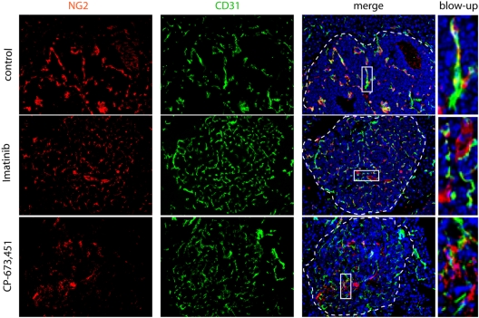
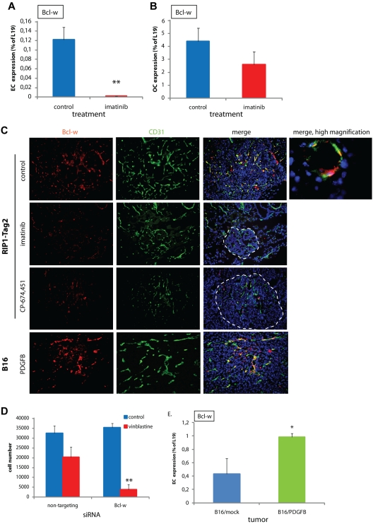
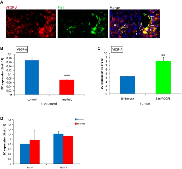
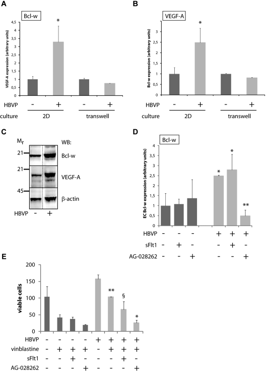
Endothelial cells (ECs) in blood vessels under formation are stabilized by the recruitment of pericytes, both in normal tissues and during angiogenesis in pathologic situations, including neoplasia. In the tumor vasculature, besides supporting the functionality of blood flow, pericytes protect ECs from antiangiogenic therapies, and have thus been implicated in clinical resistance to vascular targeting drugs. However, the molecular nature of the crosstalk between pericytes and ECs is largely unchartered. Herein, we identified pericyte-induced survival signals in ECs by isolation of vascular fragments derived from tumors that had been genetically or pharmacologically engineered to be either pericyte-rich or pericyte-poor. Pericytes induced the antiapoptotic protein Bcl-w in tumor ECs both in vivo and in vitro, thereby conveying protection from cytotoxic damage. The pericyte-dependent survival signaling in ECs was consequential to enforcement of an autocrine loop involving VEGF-A expression in ECs. Through molecular and functional studies, we delineated a signal transduction pathway in ECs downstream of integrin α(v) involving activation of NF-κB as the initiating event of the protective crosstalk from pericytes. Our elucidation of pericyte-derived pro-survival signaling in tumor ECs has potentially important implications for clinical development of antiangiogenic drugs, and suggests new therapeutic targets for rational multitargeting of cancer.


Figures

Figure 1
Figure 1
Pericytes are dissociated from ECs on treatment with PDGFR inhibitors. Treatment of RIP1-Tag2 mice with imatinib or CP-673,451 induces pericyte detachment in PNET (arrows). RIP1-Tag2 mice were treated with imatinib (150 mg/kg a day; n = 10) or CP-673,451 (50 mg/kg a day; n = 5) for 5 days by oral gavage. Pictures show immunostaining for CD31 (green) and NG2 (red) on cryosections. Arrows indicate tightly associated (control) or dissociated (imatinib and CP-673,451) pericytes in the tumor vasculature. Dotted line marks tumor:exocrine pancreas boundary. Cell nuclei (DAPI), blue. Boxes indicate areas shown in higher magnification in the right panel. Original magnification, 200×. The panels are representative of at least 5 fields in 5 tissue sections taken from 5 mice.
Figure 2
Figure 2
Bcl-w is a pericyte-induced antiapoptotic protein in ECs. (A-B) Expression of Bcl-w in ECs (A) or OCs (B) comprising cancer cells, pericytes, macrophages, and infrequent cancer-associated fibroblastic cells fractionated from PNET tumors excised from RIP1-Tag2 mice that had been treated with control or imatinib, as assessed by quantitative RT-PCR (n = 10 in each group, data shown are representative for 2 independent EC isolations). **P < .01 vs control, Student t test. (C) Immunostaining of sections from PNET of RIP1-Tag2 mice or B16/PDGFB tumors for Bcl-w (red) and CD31 (green). Cell nuclei (DAPI), blue. Original magnification, 200× (for high magnification image, 630×). The panels are representative of at least 5 fields in 5 tissue sections taken from 5 mice. (D) Number of MS1 pancreatic islet ECs after siRNA-mediated knockdown of Bcl-w and treatment with control or vinblastine (50 ng/mL). A nontargeting siRNA was used as a control. Data shown are representative for 3 independent experiments. **P < .01 vs nontargeting/vinblastine, Student t test. (E) Expression of Bcl-w in isolated ECs from B16/mock and B16/PDGFB tumors assessed by quantitative RT-PCR (n = 4 in each group, data shown are representative for 2 independent EC isolations). *P < .05 vs B16/mock, Student t test.
Figure 3
Figure 3
Pericyte association regulates expression of VEGF-A in ECs. (A) VEGF-A is expressed in a subset of ECs (arrows) in PNET from RIP1-Tag2 mice, as assessed by double in situ hybridization for VEGF-A (red) and Flk1 (green). Note that VEGF-A is abundantly expressed by tumor cells, as expected (stars). Cell nuclei (DAPI), blue. Original magnification, 400×. The panel is representative of at least 5 fields in 5 tissue sections taken from 5 mice. (B) Expression of VEGF-A is decreased in ECs in PNET from imatinib-treated RIP1-Tag2 mice compared with controls, as assessed by quantitative RT-PCR. ***P < .001 vs control, Student t test. (C) Expression of VEGF-A is increased in ECs in B16/PDGFB tumors compared with B16/mock, as assessed by quantitative RT-PCR. **P < .01 vs B16/mock, Student t test. (D) Treatment of MS1 pancreatic islet ECs with 3μM imatinib did not alter the expression of VEGF-A or Bcl-w, as assessed by quantitative RT-PCR.
Figure 4
Figure 4
A coculture system reveals the functional significance of crosstalk between ECs and pericytes. (A,B) Quantitative RT-PCR analysis of Bcl-w (A) and VEGF-A (B) expression in MS1 endothelial cells cultured alone or together with HBVPs for 96 hours, either as 2D cultures allowing physical contact between ECs and pericytes or in transwell cultures where the 2 cell types are separated by a membrane with pore size 0.4 μm. Data shown represent the mean of 9 and 2 independent experiments (2D cultures and transwell cultures, respectively). *P < .05 vs MS1 mono-culture. (C) Western blot (WB) analysis of Bcl-w and VEGF-A in cell lysates from MS1 endothelial cells cultured alone or together with HBVPs for 96 hours. Mr indicates molecular weight (kDa). Western blot for β-actin was used as a loading control. Dotted line marks the removal of one irrelevant lane from the original photograph. (D) Quantitative RT-PCR analysis of Bcl-w expression in MS1 endothelial cells cultured alone or together with HBVPs for 96 hours with the addition of the VEGF inhibitors sFlt1 (a ligand trap acting extracellularly) or AG-028262 (an intracellular inhibitor blocking VEGF signaling via its receptor tyrosine kinases). *P < .05 vs MS1 mono-culture; **P < .01 vs MS1/HBVP coculture. (E) Quantification of the number of viable MS1 endothelial cells cultured alone or together with HBVPs for 96 hours with the addition of the cytotoxic drug vinblastine (50 ng/mL) and the VEGF inhibitors sFlt1 (blocking only extracellular sources of VEGF-A) or AG-028262 (blocking both extra- and intracellular sources of VEGF-A). *P < .05 vs vinblastine-treated coculture; **P < .01 vs vinblastine-treated mono-culture; and §, not statistically significant vs vinblastine-treated coculture. HBVP indicates human brain vascular pericytes.
Figure 5
Figure 5
Pericyte-induced expression of survival genes in ECs requires NF-κB activity. (A,B) Expression of target genes of the transcription factor NF-κB was modulated in ECs from tumors in a pericyte-dependent manner, as assessed by quantitative RT-PCR on purified ECs from PNET from RIP1-Tag2 mice treated with control or imatinib (A), and from B16/mock or B16/PDGFB tumors (B). *P < .05 vs control; *P < .01 vs control. (C,D) The NF-κB inhibitor TPCK (3μM) reduced the basal expression of Bcl-w and VEGF-A in MS1 ECs (D) and sensitized MS1 cells to the action of vinblastine (50 ng/mL; D). *P < .05 vs DMSO; **P < .01 vs vinblastine; ***P < .001 vs control. (E,F) Pericytes are unable to modulate the expression of Bcl-w (E) or VEGF-A (F) in ECs on expression of the IκB-α super-repressor protein in MS1 cells. ***P < .001 vs coculture. HBVP indicates human brain vascular pericytes.
Figure 6
Figure 6
The crosstalk between pericytes and ECs is mediated by integrin αv activity. (A,B) Quantitative RT-PCR analysis of Bcl-w (A) and VEGF-A (B) expression in MS1 endothelial cells cultured alone or together with HBVPs for 96 hours in the presence or absence of neutralizing antibodies against integrin αv. Data shown are representative of 2 independent experiments. **P < .01 vs MS1/HBVP coculture. (C) Quantification of the number of viable MS1 endothelial cells cultured alone or together with HBVPs for 96 hours with the addition of the cytotoxic drug vinblastine (50 ng/mL) in the presence or absence of neutralizing antibodies against integrin αv. **P < .01 vs vinblastine coculture. (D) Immunostaining of tissue sections from PNET of RIP1-Tag2 mice for the integrin αv ligand vitronectin (green) and the pericyte marker PDGFRβ (red). Cell nuclei (DAPI), blue. Original magnification, 400×. The panels are representative of at least 5 fields in 5 tissue sections taken from 5 mice. (E,F) Quantitative RT-PCR analysis of the expression of vitronectin in nonendothelial cells of PNET from RIP1-Tag2 mice treated or not with imatinib (E), and of B16/mock or B16/PDGFB tumors (F). *P < .05 vs B16/mock; ***P < .001 vs control. (G) Quantitative PCR analysis of expression of Bcl-w and VEGF-A in MS1 cells cultured on plastic or vitronectin. **P < .01 vs plastic.
Figure 7
Figure 7
A model of pericyte-induced EC survival signaling. Endothelial cells and pericytes communicate through the reciprocal exchange of growth factors. PDGF-BB produced by ECs acts to recruit pericytes along growing vascular sprouts. In turn, pericytes confer survival advantage to ECs through PDGF-dependent secretion of vitronectin, (1) which through an integrin αv (2) and NF-κB (3) mediated signaling pathway results in up-regulation of intracrine VEGF-A signaling (4) and Bcl-w expression. (5) Previously described survival factors induced by the paracrine action of pericyte- or tumor cell-derived VEGF-A, such as survivin, Bcl-2, and XIAP, are presumably sensitive to the action of both intra- and extra-cellularly acting VEGFR inhibitors (AG-028262 and sFlt1, respectively), whereas the pericyte-induced autocrine signaling by VEGF-A is only sensitive to intra-cellularly acting inhibitors.

Comment in

References

    1. Pietras K, Ostman A. Hallmarks of cancer: interactions with the tumor stroma. Exp Cell Res. 2010;316(8):1324–1331. - PubMed
    1. Kerbel RS. Tumor angiogenesis. N Engl J Med. 2008;358(19):2039–2049. - PMC - PubMed
    1. Ebos JM, Lee CR, Kerbel RS. Tumor and host-mediated pathways of resistance and disease progression in response to antiangiogenic therapy. Clin Cancer Res. 2009;15(16):5020–5025. - PMC - PubMed
    1. Casanovas O, Hicklin DJ, Bergers G, Hanahan D. Drug resistance by evasion of antiangiogenic targeting of vegf signaling in late-stage pancreatic islet tumors. Cancer Cell. 2005;8(4):299–309. - PubMed
    1. Paez-Ribes M, Allen E, Hudock J, et al. Antiangiogenic therapy elicits malignant progression of tumors to increased local invasion and distant metastasis. Cancer Cell. 2009;15(3):220–231. - PMC - PubMed

Publication types

MeSH terms