. 2011 Aug 9;108(32):13118-23.
doi: 10.1073/pnas.1104614108. Epub 2011 Jul 21.

Pathway and mechanism of drug binding to G-protein-coupled receptors

Affiliations

Pathway and mechanism of drug binding to G-protein-coupled receptors

Ron O Dror et al. Proc Natl Acad Sci U S A. .

Abstract

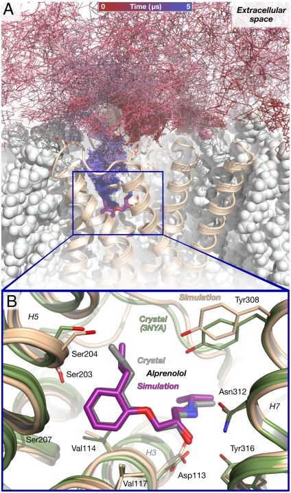
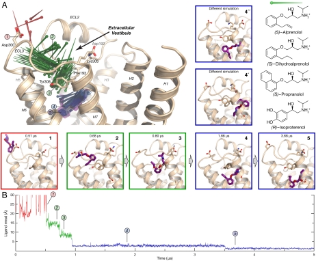
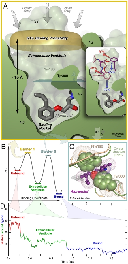
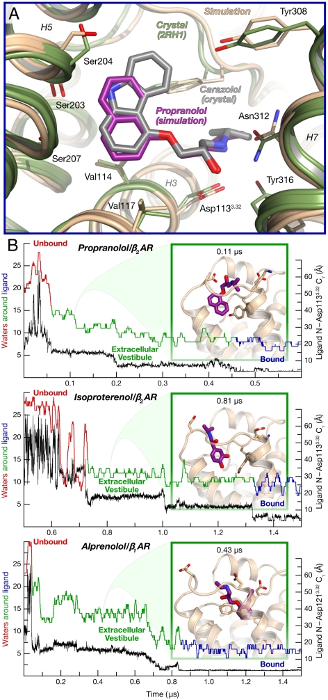
How drugs bind to their receptors--from initial association, through drug entry into the binding pocket, to adoption of the final bound conformation, or "pose"--has remained unknown, even for G-protein-coupled receptor modulators, which constitute one-third of all marketed drugs. We captured this pharmaceutically critical process in atomic detail using the first unbiased molecular dynamics simulations in which drug molecules spontaneously associate with G-protein-coupled receptors to achieve final poses matching those determined crystallographically. We found that several beta blockers and a beta agonist all traverse the same well-defined, dominant pathway as they bind to the β(1)- and β(2)-adrenergic receptors, initially making contact with a vestibule on each receptor's extracellular surface. Surprisingly, association with this vestibule, at a distance of 15 Å from the binding pocket, often presents the largest energetic barrier to binding, despite the fact that subsequent entry into the binding pocket requires the receptor to deform and the drug to squeeze through a narrow passage. The early barrier appears to reflect the substantial dehydration that takes place as the drug associates with the vestibule. Our atomic-level description of the binding process suggests opportunities for allosteric modulation and provides a structural foundation for future optimization of drug-receptor binding and unbinding rates.


Conflict of interest statement

The authors declare no conflict of interest.

Figures

Fig. 1.
Fig. 1.
Alprenolol binds spontaneously to β2AR in unbiased molecular dynamics simulations, achieving the crystallographic pose. (A) The path taken by an alprenolol molecule as it diffuses about the receptor and then binds. The final, bound alprenolol is shown as a stick figure (purple carbon atoms); the protein as a tan cartoon; and the lipid bilayer as white spheres. (B) Close-up view of the simulated alprenolol pose shown in (A) superimposed on the alprenolol–β2AR crystal structure (gray ligand, green cartoon; PDB entry 3NYA). Data from simulation 1 (Table S2).
Fig. 2.
Fig. 2.
The alprenolol–β2AR binding pathway passes through several metastable states. (A) Pins indicate successive positions of an alprenolol molecule as it binds to β2AR (the pin point is at the nitrogen atom position, and the round end is at the benzene ring center). Alprenolol moves from bulk solvent (red, pose 1), into the extracellular vestibule (green, poses 2 and 3), and finally into the binding pocket (blue, poses 4 and 5). Pose 5 matches the crystallographic pose (Fig. 1B), whereas pose 4 and two poses observed in a different simulation (4′ and 4′′) represent alternative, metastable poses in the binding pocket. The structures of ligands used in this study are shown at right. (B) Rmsd of alprenolol in simulation from the alprenolol-β2AR crystal structure, calculated after aligning on protein binding pocket Cα atoms (SI Text). Poses 4′ and 4′′ from simulation 3; remaining data from simulation 1.
Fig. 3.
Fig. 3.
Alprenolol must traverse two primary energetic barriers to bind to β2AR. (A) The first and largest barrier precedes association with the extracellular vestibule. Alprenolol molecules for which the ring center has crossed the “50% binding probability” surface (from bulk solvent into the extracellular vestibule; above to below in this image) bind more often than they escape back into the bulk. (Inset) In repeated simulations initialized with alprenolol already in the vestibule, the ligand usually proceeded into the binding pocket. Binding/escape percentages are shown. (B) Schematic free-energy landscape for binding. (C) Phe193ECL2 and Tyr3087.35 must separate for alprenolol to move from the extracellular vestibule into the binding pocket. View plane is indicated in light blue in (A). (D) Alprenolol loses more than half of its hydration shell as it enters the extracellular vestibule, and most of the rest as it enters the binding pocket (SI Text). Data from simulation 1.
Fig. 4.
Fig. 4.
Other drug–receptor pairs follow the same binding pathway. (A) Bound pose of propranolol (purple), superimposed on the carazolol–β2AR crystal structure (PDB entry 2RH1, gray) (data from simulation 15). (B) Distance to binding pocket (ligand nitrogen atom to Asp3.32 Cγ) and ligand hydration for propranolol and isoproterenol binding to β2AR, and for alprenolol binding to β1AR (data from simulations 13, 16, and 21). The insets illustrate a typical extracellular-vestibule-bound state for each simulation.

References

    1. Weiland GA, Minneman KP, Molinoff PB. Fundamental difference between the molecular interactions of agonists and antagonists with the β-adrenergic receptor. Nature. 1979;281:114–117. - PubMed
    1. Ward WH, Holdgate GA. Isothermal titration calorimetry in drug discovery. Prog Med Chem. 2001;38:309–376. - PubMed
    1. Lu H, Tonge PJ. Drug-target residence time: Critical information for lead optimization. Curr Opin Struct Biol. 2010;14:467–474. - PMC - PubMed
    1. Swinney DC. Applications of binding kinetics to drug discovery: Translation of binding mechanisms to clinically differentiated therapeutic responses. Pharmaceut Med. 2008;22:23–34.
    1. Baldwin AJ, Kay LE. NMR spectroscopy brings invisible protein states into focus. Nat Chem Biol. 2009;5:808–814. - PubMed

MeSH terms

Substances