. 2011 Aug 9;108(32):13083-8.
doi: 10.1073/pnas.1104984108. Epub 2011 Jul 21.

Structural basis for alginate secretion across the bacterial outer membrane

Affiliations

Structural basis for alginate secretion across the bacterial outer membrane

John C Whitney et al. Proc Natl Acad Sci U S A. .

Abstract

Pseudomonas aeruginosa is the predominant pathogen associated with chronic lung infection among cystic fibrosis patients. During colonization of the lung, P. aeruginosa converts to a mucoid phenotype characterized by the overproduction of the exopolysaccharide alginate. Secretion of newly synthesized alginate across the outer membrane is believed to occur through the outer membrane protein AlgE. Here we report the 2.3 Å crystal structure of AlgE, which reveals a monomeric 18-stranded β-barrel characterized by a highly electropositive pore constriction formed by an arginine-rich conduit that likely acts as a selectivity filter for the negatively charged alginate polymer. Interestingly, the pore constriction is occluded on either side by extracellular loop L2 and an unusually long periplasmic loop, T8. In halide efflux assays, deletion of loop T8 (ΔT8-AlgE) resulted in a threefold increase in anion flux compared to the wild-type or ΔL2-AlgE supporting the idea that AlgE forms a transport pathway through the membrane and suggesting that transport is regulated by T8. This model is further supported by in vivo experiments showing that complementation of an algE deletion mutant with ΔT8-AlgE impairs alginate production. Taken together, these studies support a mechanism for exopolysaccharide export across the outer membrane that is distinct from the Wza-mediated translocation observed in canonical capsular polysaccharide export systems.


Conflict of interest statement

The authors declare no conflict of interest.

Figures

Fig. 1.
Fig. 1.
Overall structure of AlgE. (A) Side view of AlgE shown as a cartoon representation. Conserved residues Phe187 and Tyr190 that extend the hydrophobic surface area above Glu166 and Arg150 of strands S6 and S5, respectively, are shown in blue. The calcium ion bound to loop L1 is shown in gray. (B) AlgE viewed from the extracellular space. Loops L2, L3, L7, and T8 are shown in cyan, black, blue, and orange, respectively. The asterisk indicates a region of L2 where no interpretable electron density is observed in the structure. (C) Slab view of AlgE showing occlusion of the pore constriction by loops L2 and T8.
Fig. 2.
Fig. 2.
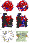
AlgE contains an electropositive pore constriction. (A and B) Electrostatic surface representation (contoured from +10 to -10 kT/e) with (left) and without (right) occluding loops L2 and T8 as seen from the extracellular space (A) and as a slab view from the plane of the outer membrane (B). Positive and negative charges are colored blue and red, respectively. (C) Stick representation of the pore-forming residues as viewed from the extracellular space. The 2Fo-Fc electron density map is contoured at 1.2σ and displayed as a gray mesh. Carbon, nitrogen, and oxygen atoms are colored yellow, blue, and red, respectively. (D) Side view of the pore constriction showing a surface representation of the interior of the pore generated using HOLLOW (45).
Fig. 4.
Fig. 4.
Conservation of the OprD-like fold. (A) Superposition of AlgE (yellow) with OprD-family member P. aeruginosa OpdK (red) viewed along the plane of the outer membrane (left) and from the extracellular space (right). (B) Structural conservation of pore forming loops L3 and L7 between AlgE (left) and OpdK (right) shown from the extracellular space (top) and periplasm (bottom). Structures are shown as surface representations with L3 and L7 colored in black and blue, respectively. For both proteins, extracellular loops that obstruct the view of the barrel interior have been removed for clarity.
Fig. 5.
Fig. 5.
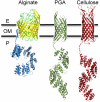
Exopolysaccharide secretion. Cartoon representation of the outer membrane proteins involved in the export of alginate (left), and the predicted models for PGA (center) and cellulose (right). AlgE and AlgK are colored in yellow and blue, respectively. AlgK is anchored in the outer membrane by its lipid anchor (blue squiggly line). E. coli PgaA and BcsC are colored in green and red, respectively. For each protein there is a variable length linker region between the lipid anchor or porin domain, and TPR superhelix (dashed line). Horizontal lines indicate the approximate boundaries of the outer membrane (OM), separating the periplasm (P), and extracellular space (E).
Fig. 3.
Fig. 3.
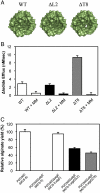
Periplasmic loop T8 occludes the pore constriction. (A) Molecular surface representation of AlgE, AlgEΔL2, and AlgEΔT8. (B) Quantification of halide efflux rates for AlgE, AlgEΔL2, and AlgEΔT8 in the absence or presence of 1 mM di-mannuronic acid (MM). All efflux rates are the mean of four independent measurements carried out from at least two separate reconstitutions of purified protein. Flux rates from empty (no protein) liposomes were subtracted from all measurements. (C) Overall amount of alginate production in P. aeruginosa PDO300(MCS-5), PDO300ΔalgE(MCS-5), PDO300ΔalgE(MCS-5:algE), PDO300ΔalgE(MCS-5:algEΔL2), and PDO300ΔalgE(MCS-5:algEΔT8). The amount of alginate produced is the mean of three independent replicates. The error bars in (B) and (C) represent the standard errors (SEM).

References

    1. Gacesa P. Bacterial alginate biosynthesis—recent progress and future prospects. Microbiology. 1998;144:1133–1143. - PubMed
    1. Ramsey DM, Wozniak DJ. Understanding the control of Pseudomonas aeruginosa alginate synthesis and the prospects for management of chronic infections in cystic fibrosis. Mol Microbiol. 2005;56:309–322. - PubMed
    1. Parsek MR, Singh PK. Bacterial biofilms: an emerging link to disease pathogenesis. Annu Rev Microbiol. 2003;57:677–701. - PubMed
    1. Lam J, Chan R, Lam K, Costerton JW. Production of mucoid microcolonies by Pseudomonas aeruginosa within infected lungs in cystic fibrosis. Infect Immun. 1980;28:546–556. - PMC - PubMed
    1. Baltimore RS, Shedd DG. The role of complement in the opsonization of mucoid and non-mucoid strains of Pseudomonas aeruginosa. Pediatr Res. 1983;17:952–958. - PubMed

Associated data

LinkOut - more resources