. 2012 Jan;46(1):14-22.
doi: 10.1165/rcmb.2010-0506OC.

Endothelin-1, the unfolded protein response, and persistent inflammation: role of pulmonary artery smooth muscle cells

Affiliations

Endothelin-1, the unfolded protein response, and persistent inflammation: role of pulmonary artery smooth muscle cells

Michael E Yeager et al. Am J Respir Cell Mol Biol. 2012 Jan.

Abstract

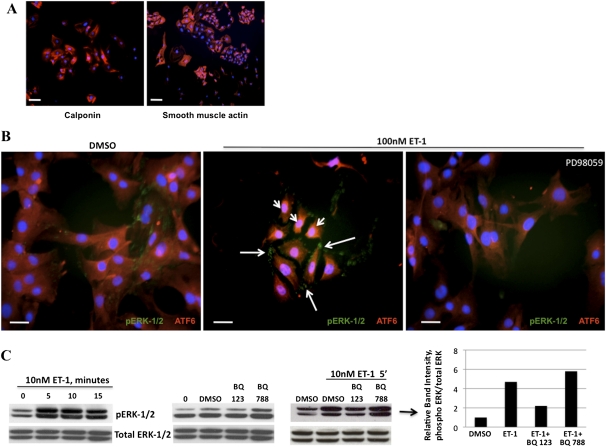
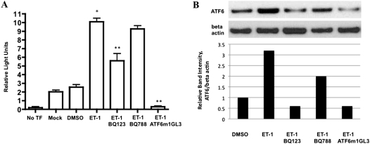
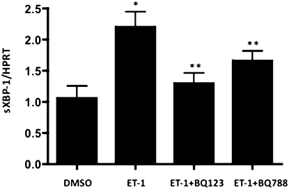
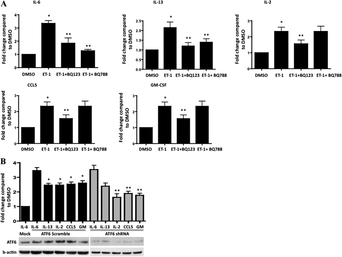
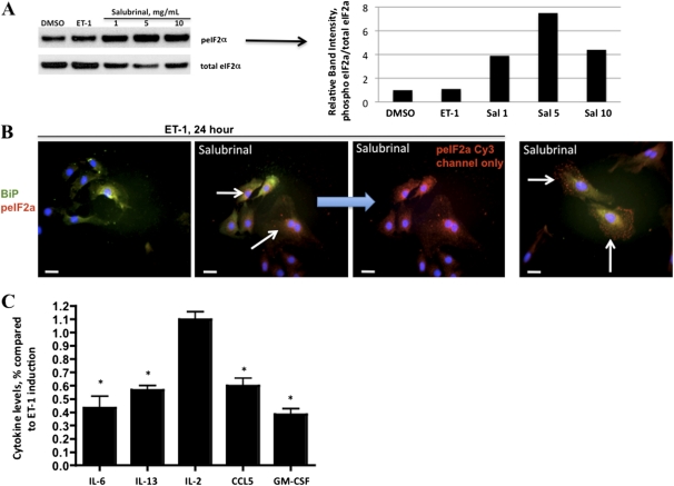
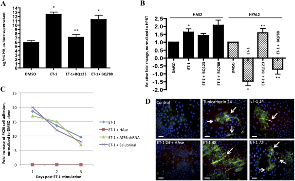
Endothelin-1 is a potent vasoactive peptide that occurs in chronically high levels in humans with pulmonary hypertension and in animal models of the disease. Recently, the unfolded protein response was implicated in a variety of diseases, including pulmonary hypertension. In addition, evidence is increasing for pathological, persistent inflammation in the pathobiology of this disease. We investigated whether endothelin-1 might engage the unfolded protein response and thus link inflammation and the production of hyaluronic acid by pulmonary artery smooth muscle cells. Using immunoblot, real-time PCR, immunofluorescence, and luciferase assays, we found that endothelin-1 induces both a transcriptional and posttranslational activation of the three major arms of the unfolded protein response. The pharmacologic blockade of endothelin A receptors, but not endothelin B receptors, attenuated the observed release, as did a pharmacologic blockade of extracellular signal-regulated kinases 1 and 2 (ERK-1/2) signaling. Using short hairpin RNA and ELISA, we observed that the release by pulmonary artery smooth muscle cells of inflammatory modulators, including hyaluronic acid, is associated with endothelin-1-induced ERK-1/2 phosphorylation and the unfolded protein response. Furthermore, the synthesis of hyaluronic acid induced by endothelin-1 is permissive for persistent THP-1 monocyte binding. These results suggest that endothelin-1, in part because it induces the unfolded protein response in pulmonary artery smooth muscle cells, triggers proinflammatory processes that likely contribute to vascular remodeling in pulmonary hypertension.


Figures

Figure 1.
Figure 1.
Endothelin-1 (ET-1) induces the phosphorylation of extracellular signal–regulated kinase 1 and 2 (ERK-1/2) by pulmonary artery smooth muscle cells (PASMCs) and activation of the activating transcription factor 6 (ATF6) arm of the unfolded protein response (UPR). (A) Fluorescence microscopy of isolated PASMCs (calponin or smooth muscle actin = Cy3, 4',6-diamidino-2-phenylindole [DAPI] nuclei). (B) Within 5 minutes, ET-1 induced the rapid nuclear localization of the UPR protein ATF6 (arrowheads, Cy3), and also caused the phosphorylation (p) of ERK-1/2 (arrows, Alexa 488), both of which could be blocked by the mitogen activated protein kinase inhibitor PD98059. (C) Immunoblot time-course analysis of the phosphorylation of ERK-1/2 induced by ET-1, which peaked at 5–10 minutes. The phosphorylation of ERK-1/2 was blocked by the preincubation of PASMCs with the endothelin A receptor (ETA) receptor antagonist BQ123, but not the endothelin B receptor (ETB) receptor antagonist BQ788. Bar = 10 μm.
Figure 2.
Figure 2.
ET-1 increases the production of ATF6 in PASMCs. Rat PASMCs were transfected with 0.1 μg of one of two luciferase reporter genes: ATF6α binding-site reporter gene ATF6GL3, or the nonresponsive ATF6α mutant site reporter ATF6 m1GL3. PASMCs were exposed for 24 hours to 100 nM ET-1, and then the relative intensity of luciferase was measured (n = 3). ET-1 induced a significant increase in ATF6-driven luciferase activity compared with control samples, the mutant ATF6 reporter m1GL3, and DMSO alone (No TF, empty cells; Mock, transfection with empty vector; DMSO, reporter plasmid transfection without stimulation by ET-1). (B) Densitometry of ATF6 Western blot of protein lysates under identical conditions as in A. *P < 0.05 compared with no TF. **P < 0.05 compared with ET-1–induced activation of ATF6 reporter.
Figure 3.
Figure 3.
ET-1 increases splicing of X-box binding protein 1 (XBP-1) in PASMCs. ET-1 activates the inositol requiring enzyme 1 (IRE-1)/XBP-1 arm of the UPR, as evidenced by a twofold increase in the spliced form of XBP-1 (sXBP-1), normalized to the housekeeping gene HPRT. This effect is inhibited by ETA antagonism (BQ123), and to a lesser extent, by ETB antagonism (BQ788). *P < 0.05, compared with DMSO. **P < 0.05, compared with ET-1–induced concentration of sXBP-1.
Figure 4.
Figure 4.
ET-1 induces the release by PASMCs of IL-6, IL-13, IL-2, CCL5, and granulocyte macrophage colony stimulating factor (GM-CSF). (A) The blockade of ETA attenuates these cytokine increases. The blockade of ETB prevents the release of IL-6 and IL-13 but not of IL-2, CCL5, or GM-CSF (*P < 0.05, compared with DMSO; **P < 0.05, compared with ET-1–induced fold change). (B) ATF6 short hairpin RNA knockdown attenuates the ET-1–mediated production of IL-2, CCL5, and GM-CSF, but not of IL-6 or IL-13. Representative immunoblots of ATF6 knockdown in rat PASMC lysates correspond to cytokine supernatant analyses (Mock, PASMCs treated with transfection reagent alone and ET-1–induced IL-6 measured in medium; GM, GM-CSF). *P < 0.05, compared with Mock. **P < 0.05, compared with ET-1 treatment with ATF6 scramble transfection.
Figure 5.
Figure 5.
ET-1 induces a release of inflammatory cytokine by PASMCs that is partly dependent on the dephosphorylation of eIF2α. (A) Immunoblot analysis of salubrinal dosing of PASMCs. Cells were pretreated overnight with salubrinal as indicated, and treated with 100 nM ET-1 and lysates taken after 24 hours. ET-1 did not increase the concentration of peIF2α, whereas salubrinal increased the concentration of peIF2α at all three doses. (B) Immunofluorescence of PASMCs treated with 100 nM ET-1 for 24 hours. With salubrinal, a pronounced increase in peIF2α is apparent (arrows, Cy3), regardless of ET-1 treatment. The localization of binding immunoglobulin protein (BiP) is not affected by either ET-1 or salubrinal. (C) Salubrinal causes decreased concentrations of IL-6, IL-13, CCL5, and GM-CSF, but not IL-2, in supernatants of PASMCs treated with ET-1. *P < 0.05, compared with induction of ET-1 as in Figure 4. Bar = 10 μm.
Figure 6.
Figure 6.
ET-1 affects biology of hyaluronic acid (HA) in rat PASMCs. (A) HA in PASMC supernatant increases in response to ET-1, and was inhibited by ETA antagonism but not by ETB antagonism. (B) Hyaluronan synthase (HAS)–2 transcripts slightly increase in response to ET-1, and this increase was independent of ETA or ETB. hyaluronidase (HYAL)–2 decreases in response to ET-1, and was reversed by the antagonism of ETA, but not of ETB. (C) Incubation for 24 hours of rat PASMCs with ET-1 induces prolonged HA-mediated THP-1 monocyte adhesion lasting 3 days. The treatment of PASMCs with ATF6 shRNA or salubrinal did not abrogate HA-mediated THP-1 adhesion. (D, from top left to right) PKH26-labeled THP-1 cells (arrows) do not adhere to untreated PASMCs, and HA is barely detectable. When PASMCs are treated with either tunicamycin or ET-1 for 24 hours, THP-1 cells adhere and HA can be detected, except if treated with hyaluronidase (HAse) before THP-1 cell incubation (bottom left). THP-1 cells persistently adhered, and HA remained detectable, at 48 and 72 hours after the stimulation by ET-1 of PASMCs (lower middle and right). *P < 0.05, compared with DMSO. **P < 0.05, compared with ET-1 alone. Bar = 10 μm.
Figure 7.
Figure 7.
Leukocytes adhere to rat pulmonary vessels rich in HA. (A and B) Peri(broncho)vascular HA was significantly more abundant in rats (B, arrows) with monocrotaline (MCT)–induced pulmonary hypertension compared with (A) control rats (biotin–HA binding protein + streptavidin–Alexa 488, with DAPI counterstain). (C) PKH26-labeled THP-1 cells did not adhere to control rat lung sections. (D) THP-1 cells (PKH26 in red, indicated by arrows) adhered to HA-rich medial and adventitial regions in MCT rat lung sections. (E) HAse pretreatment abolished THP-1 adhesion. (F) THP-1 cell adhesion quantification per bronchovascular structure was counted in five fields from five sections each, with six rats per group. Bar = 20 μm.

References

    1. Archer SL, Weir EK, Wilkins MR. Basic science of pulmonary arterial hypertension for clinicians: new concepts and experimental therapies. Circulation 2010;121:2045–2066 - PMC - PubMed
    1. Milan A, Magnino C, Veglio F. Echocardiographic indexes for the non-invasive evaluation of pulmonary hemodynamics. J Am Soc Echocardiogr 2010;23:225–239 - PubMed
    1. Hassoun PM, Mouthon L, Barberà JA, Eddahibi S, Flores SC, Grimminger F, Jones PL, Maitland ML, Michelakis ED, Morrell NW, et al. Inflammation, growth factors, and pulmonary vascular remodeling. J Am Coll Cardiol 2009;54:S10–S19 - PubMed
    1. Dorfmuller P, Humbert M, Perros F, Sanchez O, Simonneau G, Muller KM, Capron F. Fibrous remodeling of the pulmonary venous system in pulmonary arterial hypertension associated with connective tissue diseases. Hum Pathol 2007;38:893–902 - PubMed
    1. Dorfmuller P, Zarka V, Durand-Gasselin I, Monti G, Balabanian K, Garcia G, Capron F, Coulomb-Lhermine A, Marfaing-Koka A, Simonneau G, et al. Chemokine RANTES in severe pulmonary arterial hypertension. Am J Respir Crit Care Med 2002;165:534–539 - PubMed

Publication types

MeSH terms