. 2011;19(3):163-74.
doi: 10.1159/000328516. Epub 2011 Jul 22.

Resveratrol-activated AMPK/SIRT1/autophagy in cellular models of Parkinson's disease

Affiliations

Resveratrol-activated AMPK/SIRT1/autophagy in cellular models of Parkinson's disease

Yuncheng Wu et al. Neurosignals. 2011.

Abstract

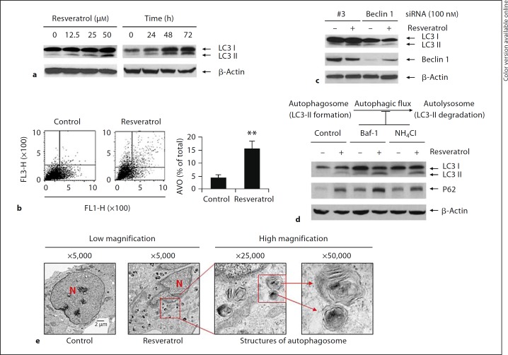
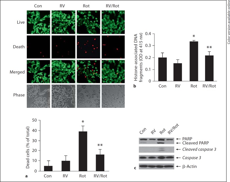
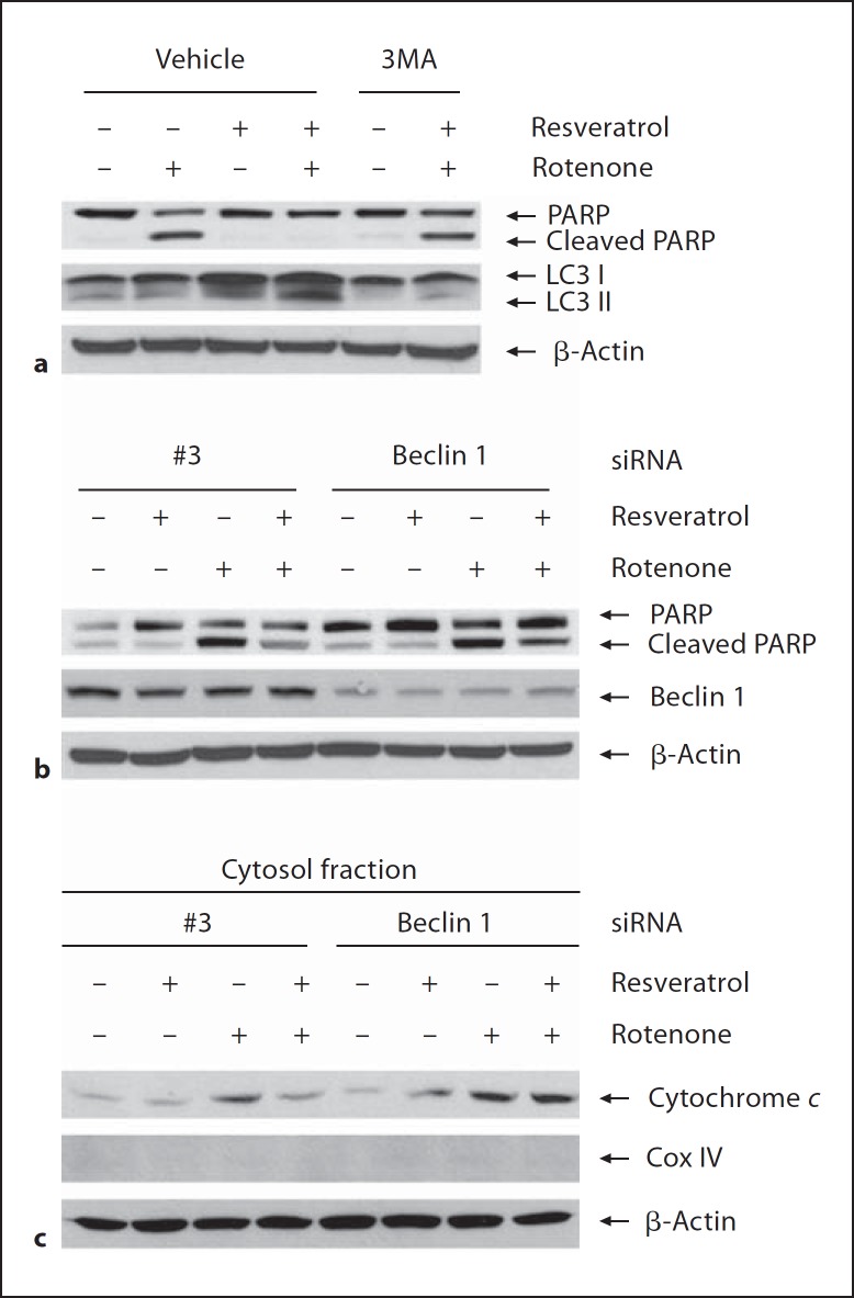
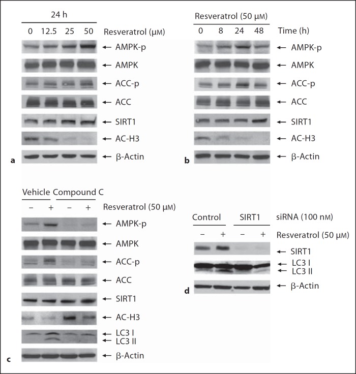
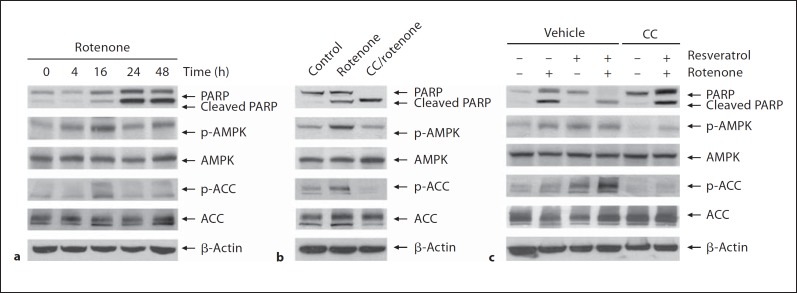
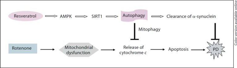
Excessive misfolded proteins and/or dysfunctional mitochondria, which may cause energy deficiency, have been implicated in the etiopathogenesis of Parkinson's disease (PD). Enhanced clearance of misfolded proteins or injured mitochondria via autophagy has been reported to have neuroprotective roles in PD models. The fact that resveratrol is a known compound with multiple beneficial effects similar to those associated with energy metabolism led us to explore whether neuroprotective effects of resveratrol are related to its role in autophagy regulation. We tested whether modulation of mammalian silent information regulator 2 (SIRT1) and/or metabolic energy sensor AMP-activated protein kinase (AMPK) are involved in autophagy induction by resveratrol, leading to neuronal survival. Our results showed that resveratrol protected against rotenone-induced apoptosis in SH-SY5Y cells and enhanced degradation of α-synucleins in α-synuclein-expressing PC12 cell lines via autophagy induction. We found that suppression of AMPK and/or SIRT1 caused decrease of protein level of LC3-II, indicating that AMPK and/or SIRT1 are required in resveratrol-mediated autophagy induction. Moreover, suppression of AMPK caused inhibition of SIRT1 activity and attenuated protective effects of resveratrol on rotenone-induced apoptosis, further suggesting that AMPK-SIRT1-autophagy pathway plays an important role in the neuroprotection by resveratrol on PD cellular models.


Figures

Fig. 1
Fig. 1
Pharmacological induction of autophagy by resveratrol. a SH-SY5Y cells were treated with resveratrol at 0, 12.5, 25, and 50 μM for 48 h or at 50 μM for 0, 24, 48 and 72 h. The protein levels of LC3 were determined by immunoblotting assay with anti-LC3 antibody. b SH-SY5Y cells were treated with resveratrol for 48 h followed by staining with acridine orange (1 μg/ml). The induction of autophagy was determined by FACScan flow cytometric analysis. The FACScan analysis profiles were shown and the top of the grid was considered as AVOs. Development of AVO was quantified and expressed as percentage of total cells. Data were mean 8 SD. Mean values were from three independent experiments. ** p < 0.01 as compared to control. c 48 h after cells were transfected with Beclin 1 siRNA or its scrambled control (#3 siRNA), cells were treated with or without resveratrol at 50 μM for additional 48 h. Cell lysates were subjected to immunoblotting assay and the protein levels of Beclin 1 and LC3 were determined with anti-Beclin 1 and anti-LC3 antibodies, respectively. d Cells were treated with 50 μM of resveratrol for 24 h. 3 h before harvest, cells were treated with Baf-1 (200 nM). Or, cells were pretreated with NH4 Cl (10 mM) overnight followed by resveratrol treatment for 24 h. Cell lysates were subjected to immunoblotting assay and the protein levels of p62 and LC3 were determined with anti-p62 and anti-LC3 antibodies. e SH-SY5Y cells were treated with resveratrol for 48 h followed by fixation. The double membrane structures of autophagy vacuous were analyzed by electron microscope under a JEM 1010 transmission electron microscope. The red square indicates autophagic vacuoles, including autophagosomes. The autophagosomes that contain intracellular contents are shown under high magnification (×50,000). N = Nucleus.
Fig. 2
Fig. 2
Alleviation of rotenone-induced injury of SH-SY5Y cells by resveratrol. SH-SY5Y cells were exposed to rotenone (10 μM) for 24 h with or without resveratrol (50 μM) pretreatment for 24 h. a Changes of cell morphology and cell survival and death were visualized using a live/dead assay. SH-SY5Y cells were stained with 1 μM calcein AM and 1.5 μM ethidium homodimer-1 for 20 min followed by the measurement of live or dead cells using fluorescence microscope. The green fluorescence, generated by calcein AM, indicates live cells, and the red fluorescence, generated by ethidium homodimer-1, indicates dead cells. Dead cells were counted for three random microscopy sections. The number of dead cells was counted and value was expressed as percentage of total cells. Data were expressed as the means ± SD. * p < 0.01 as compared to Con; ** p < 0.01 as compared to Rot. b The levels of histone-associated DNA fragmentation in the cytoplasm were quantified by ELISA assay. Data were collected from three independent experiments and expressed as means ± SD. * p < 0.01 as compared to Con; ** p < 0.05 as compared to Rot. c The whole cell lysates were subjected to immunoblotting assay to determine the protein levels of cleaved PARP and caspase-3. β-Actin was used as an equal loading of proteins. Con = Control; RV = resveratrol; Rot = rotenone.
Fig. 3
Fig. 3
Neuroprotection by resveratrol mediated through autophagy induction. a SH-SY5Y cells were pretreated with resveratrol in the absence or presence of 3MA (10 mM) for 24 h followed by exposure to rotenone for another 24 h. Apoptosis and autophagy inductions were evaluated by immunoblotting assay with anti-PARP and anti-LC3 antibodies. b SH-SY5Y cells were transfected with Beclin 1 siRNA (100 nM) or negative control #3 siRNA for 48 h followed by exposure to rotenone with or without resveratrol pretreatment. The whole cell lysates were subjected to immunoblotting assay and the apoptosis was evaluated using anti-PARP antibody. c The cytosol fraction was isolated and subjected to immunoblotting assay. The levels of cytochrome c in cytosol fraction were determined with anti-cytochrome c antibody. The purification of cytosol fraction was determined with anti-COX IV antibody.
Fig. 4
Fig. 4
Clearance of α-synuclein by resveratrol. a Wild-type, A30P or A53T α-synuclein transgenes were induced with doxycycline (1 μg/ml) in stable inducible PC12 cell lines for 48 h followed by resveratrol treatment with or without lysosomal inhibitor NH 4 Cl (10 mM) for 24 h. b Stable inducible PC12 cell lines expressing wild-type, A30P or A53T α-synuclein were transfected with Beclin 1 siRNA (100 nM) or negative control #3 siRNA. 24 h after transfection, cells were induced with doxycycline (1 μg/ml) for 36 h. The transgene expression was switched off by replacing with fresh medium followed by treatment with resveratrol for 24 h. Clearance of α-synuclein was evaluated by immunoblotting assay with anti-α-synuclein antibody.
Fig. 5
Fig. 5
Induction of autophagy by resveratrol via AMPK/SIRT1 activation. a, b SHSY5Y cells were treated with resveratrol at various concentrations for 24 h (a) or at 50 μM for various time durations (b). c SHSY5Y cells were pretreated with AMPK inhibitor compound C at 10 μ M for 3 h followed by addition of resveratrol for 24 h. d 48 h after cells were transfected with SIRT1 siRNA or its scrambled control #3 siRNA, cells were treated with or without resveratrol at 50 μM for 48 h. The whole cell lysates were subjected to immunoblotting assay and the protein levels were determined with specific antibodies indicated.
Fig. 6
Fig. 6
AMPK activation in the neuroprotection by resveratrol. a SH-SY5Y cells were exposed to rotenone (10 μM) for different time durations. b SH-SY5Y cells were treated with rotenone (10 μM) in the absence or presence of compound C (10 μM). c SH-SY5Y cells were pretreated with resveratrol (50 μM) in the presence or absence of compound C (10 μM) for 24 h followed by exposure to rotenone for 16 h. The apoptosis was evaluated by immunoblotting assay with anti-PARP antibody. The activation of AMPK was determined by measuring the protein levels of p-AMPK and p-ACC. CC = Compound C.
Fig. 7
Fig. 7
AMPK/SIRT1/autophagy-mediated neuroprotection by resveratrol. Resveratrol causes activation of AMPK/SIRT1, followed by the induction of autophagy. Induced autophagy enhances the clearance of injured mitochondria, a process termed as ‘mitophagy’, through which the protein level of cytochrome c released from injured mitochondria is decreased, leading to the reduction of rotenone-induced apoptosis. Meanwhile, induced autophagy can enhance the clearance of α-synuclein. Both the actions of resveratrol on injured mitochondria and increased α-synuclein may contribute to the neuroprotection of resveratrol on PD.

References

    1. Kopin IJ, Markey SP. MPTP toxicity, implications for research in Parkinson's disease. Annu Rev Neurosci. 1988;11:81–96. - PubMed
    1. Sherer TB, Betarberbet R, Tasta CM, Seo BB, Richardson JR, Kim JH, Miller GW, Yagi T, Matsuno-Yagi A, Greenamyre JT. Mechanism of toxicity in rotenone models of Parkinson's disease. J Neurosci. 2003;23:10756–10764. - PMC - PubMed
    1. Bonifati V, Rizzu P, van Baren MJ, Schaap O, Breedveld GJ, Krieger E, Dekker MC, Squitieri F, Ibanez P, Joosse M, van Dongen JW, Vanacore N, van Swieten JC, Brice A, Meco G, van Duijn CM, Oostra BA, Heutink P. Mutations in the DJ-1 gene associated with autosomal recessive early-onset parkinsonism. Science. 2003;299:256–259. - PubMed
    1. Valente EM, Abou-Sleiman PM, Caputo V, Muqit MM, Harvey K, Gispert S, Ali Z, Del Turco D, Bentivoglio AR, Healy DG, Albanese A, Nussbaum R, González-Maldonado R, Deller T, Salvi S, Cortelli P, Gilks WP, Latchman DS, Harvey RJ, Dallapiccola B, Auburger G, Wood NW. Hereditary early-onset Parkinson's disease caused by mutations in PINK1. Science. 2004;304:1158–1160. - PubMed
    1. West AB, Moore DJ, Biskup S, Bugayenko A, Smith WW, Ross CA, Dawson VL, Dawson TM. Parkinson's disease-associated mutations in leucine-rich repeat kinase 2 augment kinase activity. Proc Natl Acad Sci USA. 2005;102:16842–16847. - PMC - PubMed

Publication types

MeSH terms