. 2011 Jul 17;3(8):628-33.
doi: 10.1038/nchem.1087.

Tirandamycin biosynthesis is mediated by co-dependent oxidative enzymes

Affiliations

Tirandamycin biosynthesis is mediated by co-dependent oxidative enzymes

Jacob C Carlson et al. Nat Chem. .

Abstract

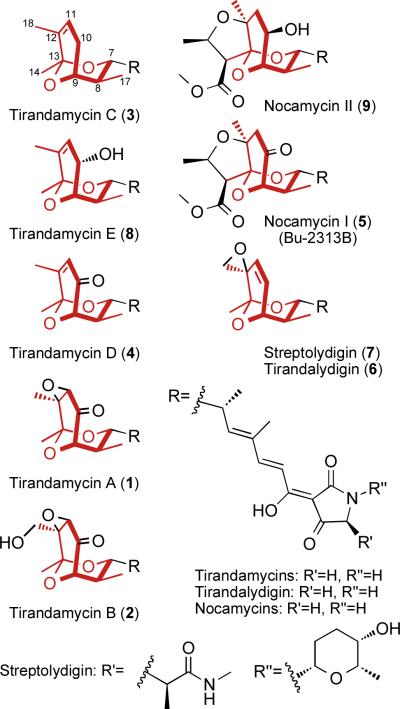
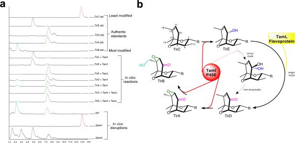
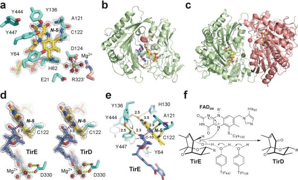
Elucidation of natural product biosynthetic pathways provides important insights into the assembly of potent bioactive molecules, and expands access to unique enzymes able to selectively modify complex substrates. Here, we show full reconstitution, in vitro, of an unusual multi-step oxidative cascade for post-assembly-line tailoring of tirandamycin antibiotics. This pathway involves a remarkably versatile and iterative cytochrome P450 monooxygenase (TamI) and a flavin adenine dinucleotide-dependent oxidase (TamL), which act co-dependently through the repeated exchange of substrates. TamI hydroxylates tirandamycin C (TirC) to generate tirandamycin E (TirE), a previously unidentified tirandamycin intermediate. TirE is subsequently oxidized by TamL, giving rise to the ketone of tirandamycin D (TirD), after which a unique exchange back to TamI enables successive epoxidation and hydroxylation to afford, respectively, the final products tirandamycin A (TirA) and tirandamycin B (TirB). Ligand-free, substrate- and product-bound crystal structures of bicovalently flavinylated TamL oxidase reveal a likely mechanism for the C10 oxidation of TirE.


Figures

Figure 1
Figure 1
Tetramic acid natural products bearing a bicyclic ketal moiety (red) with varying degrees of oxidative modification. Tirandamycin E is described within this study; all other compounds have been previously reported,. Nocamycin I and Bu-2313B are synonymous.
Figure 2
Figure 2
Spectral analysis of tirandamycin tailoring enzymes. (a) UV-vis absorption spectrum for purified TamI P450 enzyme in oxidized form (solid line), sodium dithionite reduced form (dotted line), and CO reduced form (dashed line); insets show CO-bound reduced difference spectra. (b) UV-vis absorption spectrum for purified TamI-RhFRED fusion. (c) Fluorescence excitation and emission spectra of purified TamL flavoprotein. (d) UV-vis absorption spectrum for purified TamL; inset shows UV-vis absorption spectrum of authentic FAD and FMN cofactors.
Figure 3
Figure 3
Elucidation of individual steps in the tirandamycin oxidative cascade. (a) in vitro reconstitution of TamI and TamL mediated oxidation steps, and metabolite profiles of Streptomyces sp. 307-9 wild type and mutant strains. Peaks were identified by comparison to authentic standards and MS detection of anticipated species. Relative zoom of the bottom three traces: wild type 4.1×, ΔtamI 1×, ΔtamL 17.5×. (b) Complete oxidative cascade scheme; the predominant TamI P450 reactions are highlighted in red with the intervening TamL oxidation reaction highlighted in yellow; the dashed arrows represent a minor activity toward the TamL-independent route. The R group is that shown in Figure 1. TamI mediates the initial hydroxylation of TirC at C-10, to form TirE, after which TamL oxidizes TirE to the ketone. Subsequent epoxidation and hydroxylation, both catalyzed by TamI, completes the cascade.
Figure 4
Figure 4
Ligand-free and substrate/product-bound TamL. (a) Catalytic and the Mg2+-binding sites in the substrate-free TamL with covalently bound FAD (yellow sticks) (PDB ID 2Y08). Residues from the same monomer are in cyan, from the symmetry-related monomer in pink. (b) Tirandamycin (blue sticks) in the active site of TamL (PDB ID 2Y3R). Residues 323–336 at the mouth of the substrate binding cleft are in pink. (c) Ribbon representation of TamL dimer formed by the green and pink monomers related by the non-crystallographic symmetry (PDB ID 2Y08). The Mg2+ atoms (spheres of matching colors) stabilize dimerization interface. (d) Interactions between the C-10 site of oxidation in TirE and the N-5 locus in FAD are highlighted in magenta dash line defining an angle with the N-5/N-10 flavin atoms of 110°. Installation of the keto group in TirD flattens the ketal ring pulling C-10 away from the N-5 atom by 0.3 Å (PDB ID 2Y3R). (e) Superimposition of the amino acid residues in the tirandamycin-bound (cyan sticks) compared to substrate-free (grey sticks) TamL. (f) Mechanism of dehydrogenation at C-10 in TirE. In all panels, oxygen atoms are in red, nitrogen in blue, sulfur in dark yellow, magnesium in green. Electron density 2Fo-Fc map (gray mesh) is contoured at 1.5 σ. Distances are in Angstroms.

References

    1. Carlson JC, Li S, Burr DA, Sherman DH. Isolation and characterization of tirandamycins from a marine-derived Streptomyces sp. J. Nat. Prod. 2009;72:2076–2079. - PMC - PubMed
    1. Royles BJL. Naturally occurring tetramic acids - structure, isolation, and synthesis. Chem. Rev. 1995;95:1981–2001.
    1. Carlson JC, et al. Identification of the tirandamycin biosynthetic gene cluster from Streptomyces sp. 307-9. ChemBioChem. 2010;11:564–572. - PMC - PubMed
    1. Omura T, Sato R. The carbon monoxide-binding pigment of liver microsomes. II. Solubilization, purification, and properties. J. Biol. Chem. 1964;239:2379–2385. - PubMed
    1. Li S, Podust LM, Sherman DH. In vitro characterization of a self-sufficient biosynthetic cytochrome P450 PikC fused to a heterologous reductase domain RhFRED. J. Am. Chem. Soc. 2007;129:12940–12941. - PMC - PubMed

Publication types

MeSH terms