Review
. 2011 Jul 22;11(8):573-87.
doi: 10.1038/nrc3078.

Chemotaxis in cancer

Affiliations
Review

Chemotaxis in cancer

Evanthia T Roussos et al. Nat Rev Cancer. .

Abstract

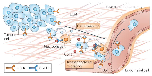
Chemotaxis of tumour cells and stromal cells in the surrounding microenvironment is an essential component of tumour dissemination during progression and metastasis. This Review summarizes how chemotaxis directs the different behaviours of tumour cells and stromal cells in vivo, how molecular pathways regulate chemotaxis in tumour cells and how chemotaxis choreographs cell behaviour to shape the tumour microenvironment and to determine metastatic spread. The central importance of chemotaxis in cancer progression is highlighted by discussion of the use of chemotaxis as a prognostic marker, a treatment end point and a target of therapeutic intervention.


Conflict of interest statement

Competing interests statement

The authors declare competing financial interests: see Web version for details.

Figures

Figure 1
Figure 1. Regulation of chemotaxis in tumour cells
a | The common cofilin activity cycle in lamellipodia and invadopodia. Different pathways regulating cofilin activity in the leading-edge protrusion (left side of the figure) and invadopodium (right side of the figure) are shown. The plasma membrane at the leading-edge protrusion is enriched with phosphatidylinositol 4,5-bisphosphate (PI(4,5)P2), and the loss of binding of cofilin to PI(4,5)P2 is the primary mechanism that is used to initiate cofilin activity at the leading edge. In invadopodia, the loss of binding of cofilin to cortactin (CTTN) is the primary mechanism used to initiate cofilin activity. New actin filaments resulting from cofilin activity support dendritic nucleation (the formation of actin branches) from the actin-related protein 2 (ARP2)–ARP3 complex, which is a common feature of both lamellipodia and invadopodia. b | The local excitation–global inhibition (LEGI) model of chemotaxis as applied to the cofilin activity cycle. Asymmetric actin polymerization (green shading) results when the asymmetric activation of cofilin (green line, which follows the concentration gradient of epidermal growth factor (EGF)) is further focused by global activation of LIM domain kinase 1 (LIMK1) (red line). LIMK1 inhibits cofilin activity globally but not fully on the side of the cell facing the chemotactic signal. The cell shown above the graph represents an example of the LEGI model, whereby a tumour cell protrudes towards a gradient of EGF that is secreted by a macrophage. As a result of this stimulation there is local asymmetric excitation of cofilin, which leads to asymmetric actin polymerization in the leading-edge protrusion (green shading), and global activation of LIMK1 within the whole cell, which results in locomotion in the direction of the arrow. ABI1, ABL-interactor 1; ARG, Abelson-related gene; DAG, diacylglycerol; GEF, guanine nucleotide exchange factor; IRSP53, insulin receptor substrate p53; MDIA1, mammalian diaphanous homologue 1; MENA, mammalian enabled homologue (also known as ENAH); NCK1, NCK adaptor protein 1; NWASP, neural Wiskott–Aldrich syndrome protein; PLCγ1, phospholipase Cγ1; ROCK, RHO-associated coiled-coil-containing protein kinase; RTK, receptor tyrosine kinase; SSH, slingshot homologue; TKS5, tyrosine kinase substrate with five SH3 domains; WAVE2, WASP family verprolin homologous protein 2.
Figure 2
Figure 2. Chemotaxis shapes the tumour microenvironment
A simplified schematic of the tumour microenvironment and the roles of chemotaxis in the processes of: immune evasion (a), angiogenesis (b) and invasion and intravasation (c) in cancer. Grey arrows indicate the gradient direction of chemotactic factors: from the cell that secretes to the cell that responds to the factor. Black arrows indicate the direction of cell migration. The dashed grey line indicates matrix fibres along which cells migrate. CSF1, colony-stimulating factor 1; CSF1R, CSF1 receptor; EGF, epidermal growth factor; EGFR, EGF receptor; PDGF, platelet-derived growth factor; TGFβ, transforming growth factor-β; VEGF, vascular endothelial growth factor; VEGFR, VEGF receptor.
Figure 3
Figure 3. Observations of streaming, intravasation and dissemination of tumour cells in mammary tumours
a | In vivo multiphoton microscopy of mammary primary tumours in mice (from REF. 5). MTLn3 rat breast adenocarcinoma cells, engineered to express a fusion protein comprised of enhanced green fluorescent protein (EGFP)–mammalian enabled homologue invasive splice variant (MENAINV; green), move in a multicellular stream towards a blood vessel (red) over 30 minutes. Scale bar = 25 μm. The white arrow indicates the direction of cell movement. The asterisk indicates the location of the blood vessel. See also Supplementary information S2 (movie). b | Immunohistochemistry of a fixed and paraffin-embedded MTLn3 primary tumour with tumour cells overexpressing MENAINV (T; pink) and F4/80-expressing macrophages (M; grey), imaged at x63 magnification (from REF. 5). Nuclear counterstain is shown in green. Scale bar = 20 μm. c | A tumour cell expressing EGFP (green) crossing the endothelium (red) of a blood vessel in a mammary tumour. Scale bar = 5 μm. Image courtesy of J. van Rheenen, J. Wyckoff and J.S.C., Albert Einstein College of Medicine. d | Photoconversion of dendra-expressing tumour cells from green to red allows the red tumour cells to be followed as they actively exit the primary tumour via blood vessels, with knowledge of their origin. e | Red photoconverted tumour cells arrive at the lung and remain there as either a disseminated non-dividing population (red) or as a dividing population (yellow). Scale bar = 25 μm. Parts d and e are reproduced, with permission, from REF. © (2011) Macmillan Publishers Ltd. All rights reserved.
Figure 4
Figure 4. Multicellular streaming of tumour cells and macrophages leading to intravasation in mammary tumours
Both streaming migration and intravasation require macrophages. In the metastatic tumour microenvironment shown, initiation of chemotaxis to epidermal growth factor (EGF) that is supplied by macrophages promotes colony-stimulating factor 1 (CSF1) production by tumour cells. Macrophages chemotax towards CSF1, resulting in relay chemotaxis between the two cell types. Relay chemotaxis results in paracrine-dependent carcinoma cell streaming and transendothelial migration. The close proximity of invasive tumour cells, macrophages and endothelial cells leads to the formation of the tumour microenvironment of metastasis (TMEM; dashed box), which has also been found as an anatomical landmark in tumour tissues from patients with breast cancer . Black arrows indicate the direction of cell migration, grey arrows indicate the gradient direction of chemotactic factors: from the cell that secretes to the cell that responds to the factor. CSF1R, CSF1 receptor; ECM, extracellular matrix; EGFR, EGF receptor.

Similar articles

Cited by

References

    1. Condeelis J, Singer RH, Segall JE. The great escape: when cancer cells hijack the genes for chemotaxis and motility. Annu Rev Cell Dev Biol. 2005;21:695–718. - PubMed
    1. McSherry EA, Donatello S, Hopkins AM, McDonnell S. Molecular basis of invasion in breast cancer. Cell Mol Life Sci. 2007;64:3201–3218. - PMC - PubMed
    1. Farrow B, Albo D, Berger DH. The role of the tumor microenvironment in the progression of pancreatic cancer. J Surg Res. 2008;149:319–328. - PubMed
    1. Condeelis J, Segall JE. Intravital imaging of cell movement in tumours. Nature Rev Cancer. 2003;3:921–930. - PubMed
    1. Roussos ET, et al. Mena invasive (MenaINV) promotes multicellular streaming motility and transendothelial migration in a mouse model of breast cancer. J Cell Sci. 2011;124:2120–2131. This study demonstrates how a MENA invasion-specific isoform promotes multicellular streaming in vivo in mouse models of breast cancer. This was the first study to define multicellular streaming migration of breast tumour cells in vivo by intravital imaging. - PMC - PubMed

Publication types