. 2011 Jul;7(7):e1002115.
doi: 10.1371/journal.ppat.1002115. Epub 2011 Jul 14.

Rotavirus stimulates release of serotonin (5-HT) from human enterochromaffin cells and activates brain structures involved in nausea and vomiting

Affiliations

Rotavirus stimulates release of serotonin (5-HT) from human enterochromaffin cells and activates brain structures involved in nausea and vomiting

Marie Hagbom et al. PLoS Pathog. 2011 Jul.

Abstract

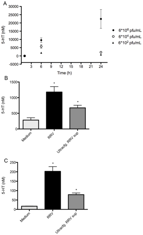
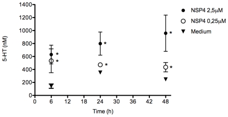
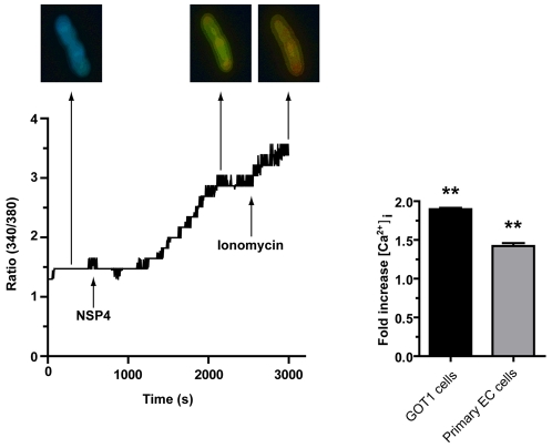
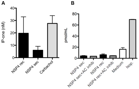
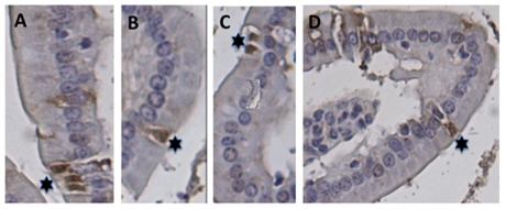
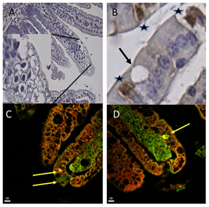
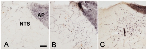
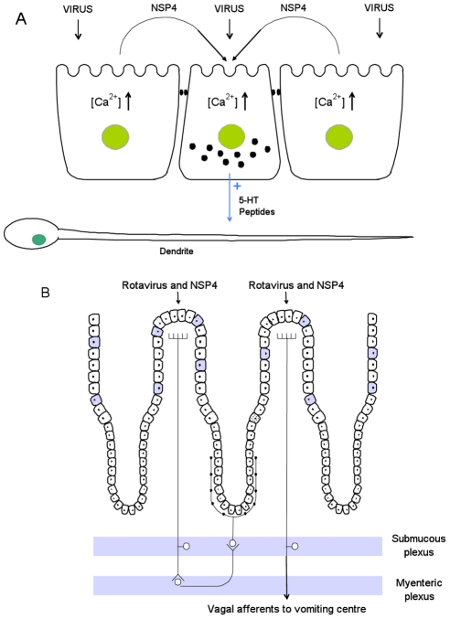
Rotavirus (RV) is the major cause of severe gastroenteritis in young children. A virus-encoded enterotoxin, NSP4 is proposed to play a major role in causing RV diarrhoea but how RV can induce emesis, a hallmark of the illness, remains unresolved. In this study we have addressed the hypothesis that RV-induced secretion of serotonin (5-hydroxytryptamine, 5-HT) by enterochromaffin (EC) cells plays a key role in the emetic reflex during RV infection resulting in activation of vagal afferent nerves connected to nucleus of the solitary tract (NTS) and area postrema in the brain stem, structures associated with nausea and vomiting. Our experiments revealed that RV can infect and replicate in human EC tumor cells ex vivo and in vitro and are localized to both EC cells and infected enterocytes in the close vicinity of EC cells in the jejunum of infected mice. Purified NSP4, but not purified virus particles, evoked release of 5-HT within 60 minutes and increased the intracellular Ca²⁺ concentration in a human midgut carcinoid EC cell line (GOT1) and ex vivo in human primary carcinoid EC cells concomitant with the release of 5-HT. Furthermore, NSP4 stimulated a modest production of inositol 1,4,5-triphosphate (IP₃), but not of cAMP. RV infection in mice induced Fos expression in the NTS, as seen in animals which vomit after administration of chemotherapeutic drugs. The demonstration that RV can stimulate EC cells leads us to propose that RV disease includes participation of 5-HT, EC cells, the enteric nervous system and activation of vagal afferent nerves to brain structures associated with nausea and vomiting. This hypothesis is supported by treating vomiting in children with acute gastroenteritis with 5-HT₃ receptor antagonists.


Conflict of interest statement

The authors have declared that no competing interests exist.

Figures

Figure 1
Figure 1. Rhesus rotavirus stimulates 5-HT release from EC tumor cells in a dose-dependent manner.
(A) Primary EC t.c. (n = 6) were infected with different concentrations of purified RRV and 5-HT secretion was measured in the cell culture supernatant at different time points by HPLC. (B) GOT1 and (C) primary EC t.c. were stimulated with RRV cell lysates (n = 3) (MOI = 1) and ultracentrifuged RRV cell lysates (n = 3) for 60 min and the 5-HT concentration was determined by ELISA. The asterisk (*) denotes statistical significance (P = 0.05; Mann Whitney test).
Figure 2
Figure 2. NSP4 induces 5-HT release in a dose- and time-dependent manner.
Primary EC t.c. were incubated with recombinant NSP4 (n = 4) at different concentrations and the 5-HT concentration in the cell medium (n = 4) was determined at different time points by HPLC. The asterisk (*) denotes statistical significance (P<0.05; Mann Whitney test).
Figure 3
Figure 3. NSP4 induces an increase in intracellular Ca2+ in human EC tumor cells.
GOT1 and primary EC t.c. were loaded with Fura-2 and Ca2+ measured before and after the addition of recombinant NSP4 (2 µM), for 40 min (GOT1 n = 8 cells, primary EC n = 10 cells). The asterisk (*) denotes statistical significance (P<0.01; Wilcoxon's rank sign test).
Figure 4
Figure 4. NSP4 activates phospholipase C mediated IP3 but not the cAMP pathway.
(A) GOT1 cells were stimulated with secretory and recombinant NSP4 (2 µM) for 60 min during accumulation of IP-one. Carbachol were used as positive control. Supernatants were then analysed for the IP3 metabolite IP-one, using a commercial ELISA (n = 3). (B) GOT1 cells were stimulated with secretory or recombinant NSP4 (2 µM) for 5 min, with or without AC inhibitor, and cAMP was measured in cell lysates of stimulated cells (n = 3) by ELISA. Isoprotenerol (Isop.) were used as positive control.
Figure 5
Figure 5. Localization of EC cells in the small intestine of mice.
(A–D) Sections of paraffin-embedded small intestine tissue from uninfected BALB/c mice were processed for immunohistochemistry with rabbit anti-chromogranin A and peroxidase-labelled goat-anti rabbit. The EC cells were identified in all segments of the small intestine. (A) Ileum, (B–D) duodenum. Stars indicate chromogranin A containing-EC cells.
Figure 6
Figure 6. Co-localization of rotavirus and 5-HT in EC tumor cells.
(A) Haematoxylin-stained jejunum of RV-infected mice. The picture shows extensive vacuoles characteristic of RV-infected enterocytes at 48 h p.i. (B) Sections of infected intestinal tissue were processed for immunohistochemistry for detection of EC cells with chromogranin A-specific antibodies. Stars indicate EC cells and the arrow identifies a vacuole in the close vicinity of an EC cell in the ileum. (C, D) Sections of jejunum intestinal tissue were stained for RV (red) and 5-HT (green). Arrows indicate EC cells with co-localization (yellow) of the virus and 5-HT.
Figure 7
Figure 7. Brain activation in response to rotavirus infection.
Coronal sections from the lower brainstem of a non-infected pup (A) and pups 48 h after RV infection (B, C). Note that many cells in the NTS of the infected animals display immunoreactivity for the activity marker Fos. AP; area postrema. Bar represents 50 µM.
Figure 8
Figure 8. Proposed disease mechanisms for rotavirus infection.
Panel A. Interplay between RV, NSP4, EC cells and enteric nervous dendrites. RV infects mature enterocytes evoking the release of NSP4, which together with invading virus, causes the release of 5-HT from EC cells. This, in turn, stimulates nervous dendrites located adjacent to the EC cells. Panel B. Physiological effects of 5-HT release from EC cells. It seems possible that the released 5-HT may activate both intrinsic afferents of the ENS as well as extrinsic afferent nerves. It is proposed that the stimulated intrinsic afferents constitute part of a nervous secretory reflex within ENS (left part of panel B), causing an intestinal fluid loss. Stimulation of extrinsic vagal afferents, activates NTS, a brain structure associated with nausea and vomiting (right part of panel B). The picture depicts the simplest nervous, secretory model that can be constructed on the basis of current experimental observations. Filled cells represent EC cells.

References

    1. Widdowson MA, Steele D, Vojdani J, Wecker J, Parashar U. Global rotavirus surveillance: determining the need and measuring the impact of rotavirus vaccines. J Infect Dis. 2009;200(Suppl 1):S1–8. - PubMed
    1. Lundgren O, Svensson L. Pathogenesis of rotavirus diarrhea. Microbes Infect. 2001;3:1145–1156. - PMC - PubMed
    1. Kordasti S, Sjovall H, Lundgren O, Svensson L. Serotonin and vasoactive intestinal peptide antagonists attenuate rotavirus diarrhoea. Gut. 2004;53:952–957. - PMC - PubMed
    1. Lundgren O, Peregrin AT, Persson K, Kordasti S, Uhnoo I, et al. Role of the enteric nervous system in the fluid and electrolyte secretion of rotavirus diarrhea. Science. 2000;287:491–495. - PubMed
    1. Ball JM, Tian P, Zeng CQ, Morris AP, Estes MK. Age-dependent diarrhea induced by a rotaviral nonstructural glycoprotein. Science. 1996;272:101–104. - PubMed

Publication types

MeSH terms