. 2011 Jul;7(7):e1002125.
doi: 10.1371/journal.ppat.1002125. Epub 2011 Jul 14.

E6 and E7 from beta HPV38 cooperate with ultraviolet light in the development of actinic keratosis-like lesions and squamous cell carcinoma in mice

Affiliations

E6 and E7 from beta HPV38 cooperate with ultraviolet light in the development of actinic keratosis-like lesions and squamous cell carcinoma in mice

Daniele Viarisio et al. PLoS Pathog. 2011 Jul.

Erratum in

Abstract

Cutaneous beta human papillomavirus (HPV) types appear to be involved in the development of non-melanoma skin cancer (NMSC); however, it is not entirely clear whether they play a direct role. We have previously shown that E6 and E7 oncoproteins from the beta HPV type 38 display transforming activities in several experimental models. To evaluate the possible contribution of HPV38 in a proliferative tissue compartment during carcinogenesis, we generated a new transgenic mouse model (Tg) where HPV38 E6 and E7 are expressed in the undifferentiated basal layer of epithelia under the control of the Keratin 14 (K14) promoter. Viral oncogene expression led to increased cellular proliferation in the epidermis of the Tg animals in comparison to the wild-type littermates. Although no spontaneous formation of tumours was observed during the lifespan of the K14 HPV38 E6/E7-Tg mice, they were highly susceptible to 7,12-dimethylbenz(a)anthracene (DMBA)/12-0-tetradecanoylphorbol-13-acetate (TPA) two-stage chemical carcinogenesis. In addition, when animals were exposed to ultraviolet light (UV) irradiation, we observed that accumulation of p21(WAF1) and cell-cycle arrest were significantly alleviated in the skin of Tg mice as compared to wild-type controls. Most importantly, chronic UV irradiation of Tg mice induced the development of actinic keratosis-like lesions, which are considered in humans as precursors of squamous cell carcinomas (SCC), and subsequently of SCC in a significant proportion of the animals. In contrast, wild-type animals subjected to identical treatments did not develop any type of skin lesions. Thus, the oncoproteins E6 and E7 from beta HPV38 significantly contribute to SCC development in the skin rendering keratinocytes more susceptible to UV-induced carcinogenesis.


Conflict of interest statement

The authors have declared that no competing interests exist.

Figures

Figure 1
Figure 1. HPV38 E6 and E7 expression in Tg mice.
(A) Schematic representation of the K14-HPV38 E6/E7 construct. (B) HPV38 E6 and E7 transcripts are differentially expressed in the epithelia of the two hemizygous Tg mouse lines 183, and 187. Total RNA was extracted from the ear, the skin, tongue, esophagus, and liver. After preparation of cDNA, E6 and E7 expression was determined by RT-qPCR and normalized towards the expression level of GAPDH. The data shown in the Figures are the means ±SD of three independent experiments. In each experiment the 187 ear data is set to 1 and the other values are consequently resized.
Figure 2
Figure 2. Histological analysis of skin specimens from wild-type FVB/N and Tg mouse lines.
Representative pictures (original magnification 40×) of HE-stained sections of paraffin-embedded tissues are shown: (A) ear (left panel) and dorsal skin (right panel) of wild-type FVB/N and Tg mice of the lines 183, and 187. (B) Dysplastic ear skin of K14 HPV38 E6/E7-Tg mice.
Figure 3
Figure 3. Analysis of cellular proliferation in the ear and dorsal skin of wild-type and transgenic mice.
(A and B, top panels) Representative pictures of Ki-67 immunostained sections of paraffin-embedded ear skin and dorsal skin from wild-type (FVB/N) and Tg animals (lines 183 and 187). (A and B, lower panels) Quantification of Ki-67-positive cells (brownish signal) in wild-type and Tg epidermis was done by counting 400 hematoxylin-stained cells under 40× magnification in 4 different fields of epidermis. Differences between the Ki-67-positive cells in the HPV38 E6/E7 Tg mice (lines 183 and 187) versus the FVB/N mice were statistically significant as determined by Student's t-test with Welch correction for unequal standard deviation.
Figure 4
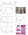
Figure 4. Tumour burden in wild-type and transgenic mice after DMBA/TPA treatment.
(A) Schematic diagram of the initiation and promotion protocol using DMBA as initiator and TPA as promoter of the two-stage skin carcinogenesis approach. (B) Tumour incidence. Percentage of animals with skin tumours in the group of FVB/N wild-type and Tg cohorts of lines 183 and 187. Skin tumour formation was recorded each week until the end of the experiment in week 24 after the beginning of promotion. The difference between the curves of control and transgenic mice is statistically significant (p<0.0001 determined by logrank test for group data). (C) Representative pictures of dorsal skin from wild-type FVB/N and Tg mice 10 weeks after the beginning of tumour promotion. (D) Survival curves for DMBA/TPA-treated wild-type FVB/N and Tg animal cohorts of lines 183 and 187. Mice were sacrificed when putative SCC skin lesions reached the size of 15 mm in diameter. The difference between the curves of control and transgenic mice is statistically significant (p<0.0001 determined by logrank test for group data). (E) Tumour multiplicity. Maximum number of tumours per animal in the groups of wild-type and Tg lines. The number of tumours was recorded every week. Differences between the tumour multiplicity in the group of wild-type and Tg lines are statistically significant (control versus line 183, p<0.001; control versus line 187, p<0.001 as determined by Wilcoxon rank sum test). (F) Incidence of cutaneous SCC in the group of wild-type and transgenic mice. SCC in the sacrificed wild-type and Tg animals were confirmed by histological analyses. The difference between the curves of control and transgenic mice is statistically significant (p<0.0001 determined by logrank test for group data). (G) Representative pictures of HE-stained skin lesions from wild-type (FVB/N) and K14 HPV38 E6/E7-Tg mice (lines 183 and 187) collected after 10 weeks of chemical carcinogens treatment (original magnification 5×). Magnified areas are shown in the right panels.
Figure 5
Figure 5. p21WAF1 and Ki-67 levels in the skin of wild-type and K14 HPV38 E6/E7-Tg mice after UVB irradiation.
Wild-type and Tg animals were irradiated up to 5 times as described in Materials and Methods. 24 hours after the last irradiation, mice were sacrificed and skin tissue was analyzed by immuno-histochemistry. (A) Representative Ki-67 and p21WAF1 immunostainings of skin from wild-type and Tg mice non-exposed (0×) or four time (4×) exposed to UVB. (B) Quantification of p21WAF1 and Ki-67-positive cells in skin of wild-type and Tg mice before and after UVB irradiation. The percentage of p21WAF1 and Ki-67-positive cells in the epidermis was determined as described in the legend of Figure 3. The differences between the percentages of p21WAF1 or Ki-67-positive cells in the HPV38 E6/E7 Tg mice (lines 183 and 187) versus the FVB/N non-Tg mice are statistically significant (* = p<0.05, ** = p<0,001) as determined by Student's t-test.
Figure 6
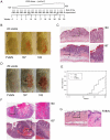
Figure 6. Tumour burden in wild-type and K14 HPV38 E6/E7-Tg animals upon UVB irradiation.
(A) Schematic diagram of the experimental procedure of long-term UVB irradiation. (B) Representative pictures of dorsal skin from wild-type FVB/N and HPV-Tg mice exposed to UVB light for 24 weeks. (C) Representative pictures of HE-stained actinic keratosis affected epidermis (AK) from K14 HPV38 E6/E7-Tg mouse lines 183 and 187 after 24 weeks of irradiation (original magnification 10×). Magnified areas are shown in the right panels. (D) Representative pictures of dorsal skin from wild-type FVB/N and HPV-Tg mice exposed to UVB light for 29 weeks. (E) Percentage of animals with skin SCC in wild-type and Tg cohorts of lines 183 and 187. Tumour formation was monitored each week until the end of the experiment in week 30 after start of treatment, and confirmed by histological analyses after sacrifice of the animals. The difference between the curves of control and transgenic mice is statistically significant (p<0,0001 determined by logrank test for group data). (F) Representative pictures of HE-stained of SCC sections (SCC) from K14 HPV38 E6/E7-Tg mouse lines 183 and 187 after 30 weeks of UVB irradiation (original magnification 5×). Magnified areas are shown in the right panels. (G) Representative pictures of HE-stained epidermis from wild-type FVB/N mice after 30 weeks of UVB irradiation (original magnification 10×). Magnified area is shown in the right panel.

Similar articles

Cited by

References

    1. Pisani P, Bray F, Parkin DM. Estimates of the world-wide prevalence of cancer for 25 sites in the adult population. Int J Cancer. 2002;97:72–81. - PubMed
    1. Ananthaswamy HN, Loughlin SM, Cox P, Evans RL, Ullrich SE, et al. Sunlight and skin cancer: inhibition of p53 mutations in UV-irradiated mouse skin by sunscreens. Nat Med. 1997;3:510–514. - PubMed
    1. Armstrong BK, Kricker A. The epidemiology of UV induced skin cancer. J Photochem Photobiol B. 2001;63:8–18. - PubMed
    1. Preston DS, Stern RS. Nonmelanoma cancers of the skin. N Engl J Med. 1992;327:1649–1662. - PubMed
    1. Boyle J, MacKie RM, Briggs JD, Junor BJ, Aitchison TC. Cancer, warts, and sunshine in renal transplant patients. A case-control study. Lancet. 1984;1:702–705. - PubMed

Publication types

MeSH terms

Substances