

High-Grade Breast Epithelioid Angiosarcoma Secondary to Radiotherapy Metastasizing to the Contralateral Lymph Node: Unusual Presentation and Potential Pitfall
- PMID: 21779229
- PMCID: PMC3132971
- DOI: 10.1159/000329323
High-Grade Breast Epithelioid Angiosarcoma Secondary to Radiotherapy Metastasizing to the Contralateral Lymph Node: Unusual Presentation and Potential Pitfall
Abstract
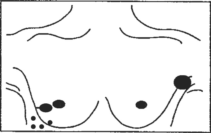
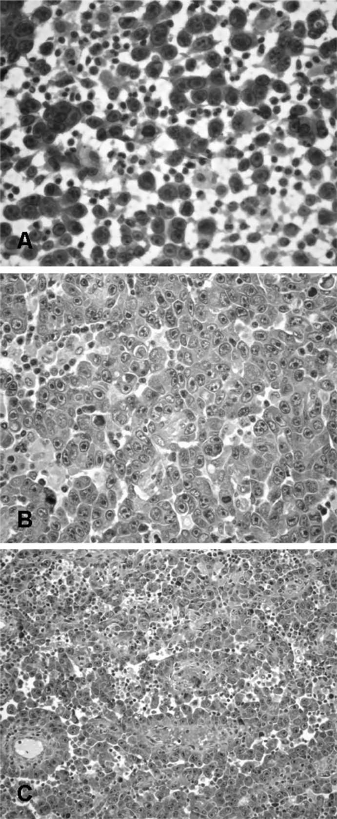
BACKGROUND: Breast angiosarcoma is a rare disease occurring as primary tumour or secondary to lymphoedema or radiotherapy. The more frequent use of breast-conserving therapy and radiotherapy for breast carcinoma explains the increasing diagnosis of these tumours. CASE REPORT: We report a case of a breast epithelioid angiosarcoma which metastasized to the contralateral axillary lymph node, occurring 4 years after breast-conserving therapy with axillary lymph node dissection and radiotherapy. The patient presented skin lesions and an axillary lump (clinically diagnosed as carcinoma relapse and lymph node metastasis). Fine-needle cytology on both lesions and a core needle biopsy of the axillary lump were carried out. Differential diagnosis included carcinoma, malignant melanoma, and angiosarcoma. Immunohistochemistry confirmed the diagnosis of angiosarcoma. CONCLUSIONS: Breast angiosarcoma is a challenge - clinically, radiologically and pathologically - and requires a high index of suspicion in susceptible patients.
Hintergrund: Angiosarkome der Brust sind selten und treten entweder als Primärtumor oder sekundär infolge von Lymphódemen oder Radiotherapie auf. Der häufiger werdende Einsatz brusterhaltender Chirurgie und Radiotherapie beim Mammakarzinom erklärt die steigende Anzahl diagnostizierter Angiosarkome der Brust.
Fallbericht: Wir berich-ten von einer Patientin, die 4 Jahre nach brusterhaltender Therapie mit axillärer Lymphknotendissektion und Radiotherapie ein epitheloides Angiosarkom der Brust mit Metas-tasierung in den kontralateralen Achsellymphknoten entwickelte. Die Patientin wurde mit Hautläsionen und einem axillären Knoten (klinisch diagnostiziert als Karzinomrezidiv und Lymphknotenmetastase) vorstellig. Es wurden Fein-nadelbiopsien beider Läsionen sowie eine Stanzbiopsie des axillären Knotens durchgeführt. Differentialdiagnostisch kamen Karzinom, malignes Melanom und Angiosarkom in Betracht. Die immunhistochemische Analyse bestätigte das Vorliegen eines Angiosarkoms.
Schlussfolgerungen: Angiosarkome der Brust sind eine Herausforderung – klinisch, radiologisch und pathologisch – und erfordern ein hohes Maß an Aufmerksamkeit bei der Untersuchung entsprechend gefährdeter Patienten.
Figures
References
-
- Brenn T, Fletcher CD. Postradiation vascular proliferations: an increasing problem. Histopathology. 2006;48:106–114. - PubMed
-
- West JG, Qureshi A, West JE, Chacon M, Sutherland ML, Haghighi B, Harrison J. Risk of angiosarcoma following breast conservation: a clinical alert. Breast J. 2005;11:115–123. - PubMed
-
- Dean CT, Jubelirer SJ, Plants BA, Welch CA. Use of radiation after breast conserving surgery (BCS) for DCIS and early invasive breast cancer at Charleston Area Medical Center (CAMC). A study of compliance with National Comprehensive Cancer Network (NCCN) guidelines. W V Med J. 2009;105:34–38. quiz39. - PubMed
-
- Goodwin A, Parker S, Ghersi D, Wilcken N. Postoperative radiotherapy for ductal carcinoma in situ of the breast. Cochrane Database Syst Rev. 2009;(4):CD000563. - PubMed
Publication types
LinkOut - more resources
Full Text Sources
