

Activation of Arabidopsis seed hair development by cotton fiber-related genes
- PMID: 21779324
- PMCID: PMC3136922
- DOI: 10.1371/journal.pone.0021301
Activation of Arabidopsis seed hair development by cotton fiber-related genes
Abstract
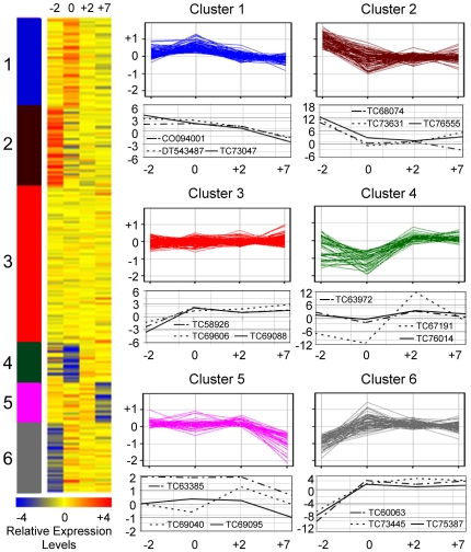
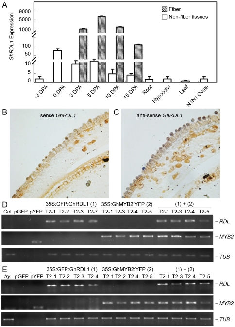
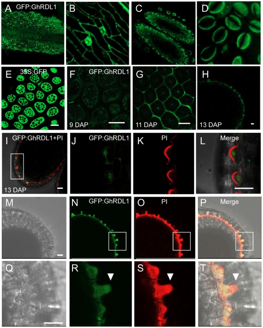
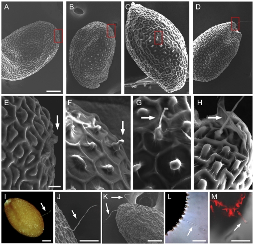
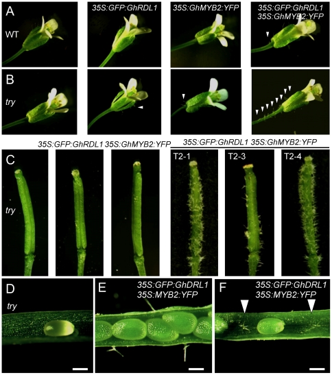
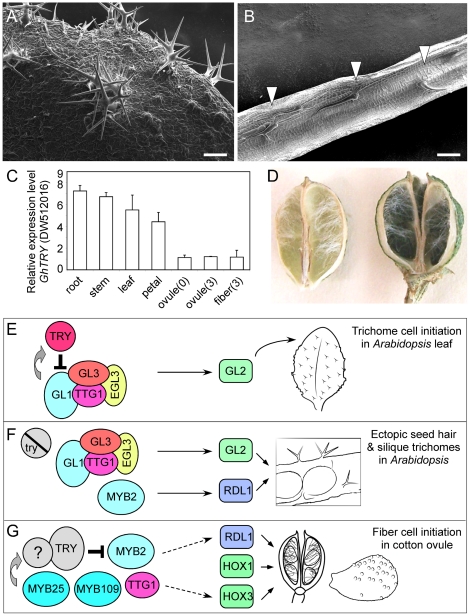
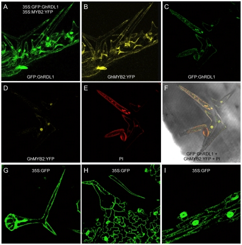
Each cotton fiber is a single-celled seed trichome or hair, and over 20,000 fibers may develop semi-synchronously on each seed. The molecular basis for seed hair development is unknown but is likely to share many similarities with leaf trichome development in Arabidopsis. Leaf trichome initiation in Arabidopsis thaliana is activated by GLABROUS1 (GL1) that is negatively regulated by TRIPTYCHON (TRY). Using laser capture microdissection and microarray analysis, we found that many putative MYB transcription factor and structural protein genes were differentially expressed in fiber and non-fiber tissues. Gossypium hirsutum MYB2 (GhMYB2), a putative GL1 homolog, and its downstream gene, GhRDL1, were highly expressed during fiber cell initiation. GhRDL1, a fiber-related gene with unknown function, was predominately localized around cell walls in stems, sepals, seed coats, and pollen grains. GFP:GhRDL1 and GhMYB2:YFP were co-localized in the nuclei of ectopic trichomes in siliques. Overexpressing GhRDL1 or GhMYB2 in A. thaliana Columbia-0 (Col-0) activated fiber-like hair production in 4-6% of seeds and had on obvious effects on trichome development in leaves or siliques. Co-overexpressing GhRDL1 and GhMYB2 in A. thaliana Col-0 plants increased hair formation in ∼8% of seeds. Overexpressing both GhRDL1 and GhMYB2 in A. thaliana Col-0 try mutant plants produced seed hair in ∼10% of seeds as well as dense trichomes inside and outside siliques, suggesting synergistic effects of GhRDL1 and GhMYB2 with try on development of trichomes inside and outside of siliques and seed hair in A. thaliana. These data suggest that a different combination of factors is required for the full development of trichomes (hairs) in leaves, siliques, and seeds. A. thaliana can be developed as a model a system for discovering additional genes that control seed hair development in general and cotton fiber in particular.
Conflict of interest statement
Figures
Similar articles
-
Functional characterization of a basic helix-loop-helix (bHLH) transcription factor GhDEL65 from cotton (Gossypium hirsutum).Physiol Plant. 2016 Oct;158(2):200-12. doi: 10.1111/ppl.12450. Epub 2016 Jun 6. Physiol Plant. 2016. PMID: 27080593
-
Control of plant trichome development by a cotton fiber MYB gene.Plant Cell. 2004 Sep;16(9):2323-34. doi: 10.1105/tpc.104.024844. Epub 2004 Aug 17. Plant Cell. 2004. PMID: 15316114 Free PMC article.
-
The MYB transcription factor GhMYB25 regulates early fibre and trichome development.Plant J. 2009 Jul;59(1):52-62. doi: 10.1111/j.1365-313X.2009.03847.x. Epub 2009 Feb 26. Plant J. 2009. PMID: 19309462
-
Updates on molecular mechanisms in the development of branched trichome in Arabidopsis and nonbranched in cotton.Plant Biotechnol J. 2019 Sep;17(9):1706-1722. doi: 10.1111/pbi.13167. Epub 2019 Jun 11. Plant Biotechnol J. 2019. PMID: 31111642 Free PMC article. Review.
-
Gene expression changes and early events in cotton fibre development.Ann Bot. 2007 Dec;100(7):1391-401. doi: 10.1093/aob/mcm232. Epub 2007 Sep 27. Ann Bot. 2007. PMID: 17905721 Free PMC article. Review.
Cited by
-
Identification and functional analysis of PIN family genes in Gossypium barbadense.PeerJ. 2022 Oct 18;10:e14236. doi: 10.7717/peerj.14236. eCollection 2022. PeerJ. 2022. PMID: 36275460 Free PMC article.
-
Functional genomics of fuzzless-lintless mutant of Gossypium hirsutum L. cv. MCU5 reveal key genes and pathways involved in cotton fibre initiation and elongation.BMC Genomics. 2012 Nov 14;13:624. doi: 10.1186/1471-2164-13-624. BMC Genomics. 2012. PMID: 23151214 Free PMC article.
-
Role of Actin Dynamics and GhACTIN1 Gene in Cotton Fiber Development: A Prototypical Cell for Study.Genes (Basel). 2023 Aug 18;14(8):1642. doi: 10.3390/genes14081642. Genes (Basel). 2023. PMID: 37628693 Free PMC article. Review.
-
Functional characterization of AGAMOUS-subfamily members from cotton during reproductive development and in response to plant hormones.Plant Reprod. 2017 Mar;30(1):19-39. doi: 10.1007/s00497-017-0297-y. Epub 2017 Feb 7. Plant Reprod. 2017. PMID: 28176007
-
Dynamic Roles for Small RNAs and DNA Methylation during Ovule and Fiber Development in Allotetraploid Cotton.PLoS Genet. 2015 Dec 28;11(12):e1005724. doi: 10.1371/journal.pgen.1005724. eCollection 2015 Dec. PLoS Genet. 2015. PMID: 26710171 Free PMC article.
References
-
- Szymanski DB, Lloyd AM, Marks MD. Progress in the molecular genetic analysis of trichome initiation and morphogenesis in Arabidopsis. Trends Plant Sci. 2000;5:214–219. - PubMed
-
- Hülskamp M. Plant trichomes: a model for cell differentiation. Nat Rev Mol Cell Biol. 2004;5:471–480. - PubMed
-
- Pesch M, Hulskamp M. One, two, three…models for trichome patterning in Arabidopsis? Curr Opin Plant Biol. 2009;12:587–592. - PubMed
-
- Esch JJ, Chen MA, Hillestad M, Marks MD. Comparison of TRY and the closely related At1g01380 gene in controlling Arabidopsis trichome patterning. Plant J. 2004;40:860–869. - PubMed
Publication types
MeSH terms
Substances
LinkOut - more resources
Full Text Sources
Other Literature Sources
Molecular Biology Databases
