Transgenic overexpression of Tcfap2c/AP-2gamma results in liver failure and intestinal dysplasia
- PMID: 21779369
- PMCID: PMC3135619
- DOI: 10.1371/journal.pone.0022034
Transgenic overexpression of Tcfap2c/AP-2gamma results in liver failure and intestinal dysplasia
Abstract
Background: The transcription factor Tcfap2c has been demonstrated to be essential for various processes during mammalian development. It has been found to be upregulated in various undifferentiated tumors and is implicated with poor prognosis. Tcfap2c is reported to impinge on cellular proliferation, differentiation and apoptosis. However, the physiological consequences of Tcfap2c-expression remain largely unknown.
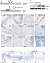
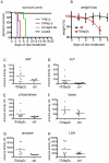
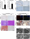
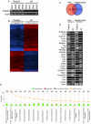
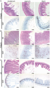
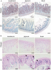
Methodology/principal findings: Therefore we established a gain of function model to analyze the role of Tcfap2c in development and disease. Induction of the transgene led to robust expression in all tissues (except brain and testis) and lead to rapid mortality within 3-7 days. In the liver cellular proliferation and apoptosis was detected. Accumulation of microvesicular lipid droplets and breakdown of major hepatic metabolism pathways resulted in steatosis. Serum analysis showed a dramatic increase of enzymes indicative for hepatic failure. After induction of Tcfap2c we identified a set of 447 common genes, which are deregulated in both liver and primary hepatocyte culture. Further analysis showed a prominent repression of the cytochrome p450 system, PPARA, Lipin1 and Lipin2. These data indicate that in the liver Tcfap2c represses pathways, which are responsible for fatty acid metabolism. In the intestine, Tcfap2c expression resulted in expansion of Sox9 positive and proliferative active epithelial progenitor cells resulting in dysplastic growth of mucosal crypt cells and loss of differentiated mucosa.
Conclusions: The transgenic mice show that ectopic expression of Tcfap2c is not tolerated. Due to the phenotype observed, iTcfap2c-mice represent a model system to study liver failure. In intestine, Tcfap2c induced cellular hyperplasia and suppressed terminal differentiation indicating that Tcfap2c serves as a repressor of differentiation and inducer of proliferation. This might be achieved by the Tcfap2c mediated activation of Sox9 known to be expressed in intestinal and hepatic stem/progenitor cell populations.
Conflict of interest statement
Figures

Similar articles
-
Transcription factor AP-2γ is a core regulator of tight junction biogenesis and cavity formation during mouse early embryogenesis.Development. 2012 Dec;139(24):4623-32. doi: 10.1242/dev.086645. Development. 2012. PMID: 23136388 Free PMC article.
-
The transcription factor TCFAP2C/AP-2gamma cooperates with CDX2 to maintain trophectoderm formation.Mol Cell Biol. 2010 Jul;30(13):3310-20. doi: 10.1128/MCB.01215-09. Epub 2010 Apr 19. Mol Cell Biol. 2010. PMID: 20404091 Free PMC article.
-
Evidence that transcription factor AP-2γ is not required for Oct4 repression in mouse blastocysts.PLoS One. 2013 May 31;8(5):e65771. doi: 10.1371/journal.pone.0065771. Print 2013. PLoS One. 2013. PMID: 23741512 Free PMC article.
-
The role of BLIMP1 and its putative downstream target TFAP2C in germ cell development and germ cell tumours.Int J Androl. 2011 Aug;34(4 Pt 2):e152-8; discussion e158-9. doi: 10.1111/j.1365-2605.2011.01167.x. Epub 2011 May 12. Int J Androl. 2011. PMID: 21564135 Review.
-
Lipin family proteins--key regulators in lipid metabolism.Ann Nutr Metab. 2015;66(1):10-8. doi: 10.1159/000368661. Epub 2014 Dec 2. Ann Nutr Metab. 2015. PMID: 25678092 Review.
Cited by
-
Recombinant Laminins Drive the Differentiation and Self-Organization of hESC-Derived Hepatocytes.Stem Cell Reports. 2015 Dec 8;5(6):1250-1262. doi: 10.1016/j.stemcr.2015.10.016. Epub 2015 Nov 25. Stem Cell Reports. 2015. PMID: 26626180 Free PMC article.
-
Human sFLT1 Leads to Severe Changes in Placental Differentiation and Vascularization in a Transgenic hsFLT1/rtTA FGR Mouse Model.Front Endocrinol (Lausanne). 2019 Mar 21;10:165. doi: 10.3389/fendo.2019.00165. eCollection 2019. Front Endocrinol (Lausanne). 2019. PMID: 30949132 Free PMC article.
-
The AP-2 Family of Transcription Factors-Still Undervalued Regulators in Gastroenterological Disorders.Int J Mol Sci. 2024 Aug 23;25(17):9138. doi: 10.3390/ijms25179138. Int J Mol Sci. 2024. PMID: 39273087 Free PMC article. Review.
-
TFAP2 transcription factors are regulators of lipid droplet biogenesis.Elife. 2018 Sep 26;7:e36330. doi: 10.7554/eLife.36330. Elife. 2018. PMID: 30256193 Free PMC article.
-
Impaired liver regeneration and lipid homeostasis in CCl4 treated WDR13 deficient mice.Lab Anim Res. 2020 Nov 13;36(1):41. doi: 10.1186/s42826-020-00076-8. Lab Anim Res. 2020. PMID: 33292732 Free PMC article.
References
-
- Williams T, Admon A, Lüscher B, Tjian R. Cloning and expression of AP-2, a cell-type-specific transcription factor that activates inducible enhancer elements. Genes Dev. 1988;2:1557–1569. - PubMed
-
- Moser M, Imhof A, Pscherer A, Bauer R, Amselgruber W, et al. Cloning and characterization of a second AP-2 transcription factor: AP-2 beta. Development (Cambridge, England) 1995;121:2779–2788. - PubMed
-
- Oulad-Abdelghani M, Bouillet P, Chazaud C, Dollé P, Chambon P. AP-2.2: a novel AP-2-related transcription factor induced by retinoic acid during differentiation of P19 embryonal carcinoma cells. Exp Cell Res. 1996;225:338–347. - PubMed
-
- Zhao F, Satoda M, Licht JD, Hayashizaki Y, Gelb BD. Cloning and characterization of a novel mouse AP-2 transcription factor, AP-2delta, with unique DNA binding and transactivation properties. J Biol Chem. 2001;276:40755–40760. - PubMed
-
- Feng W, Williams T. Cloning and characterization of the mouse AP-2 epsilon gene: a novel family member expressed in the developing olfactory bulb. Mol Cell Neurosci. 2003;24:460–475. - PubMed
Publication types
MeSH terms
Substances
LinkOut - more resources
Full Text Sources
Molecular Biology Databases
Research Materials

