. 2011;6(7):e22290.
doi: 10.1371/journal.pone.0022290. Epub 2011 Jul 14.

Structural and functional profiling of the human histone methyltransferase SMYD3

Affiliations

Structural and functional profiling of the human histone methyltransferase SMYD3

Kenneth W Foreman et al. PLoS One. 2011.

Abstract

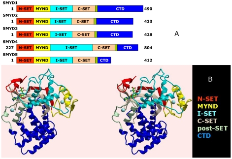
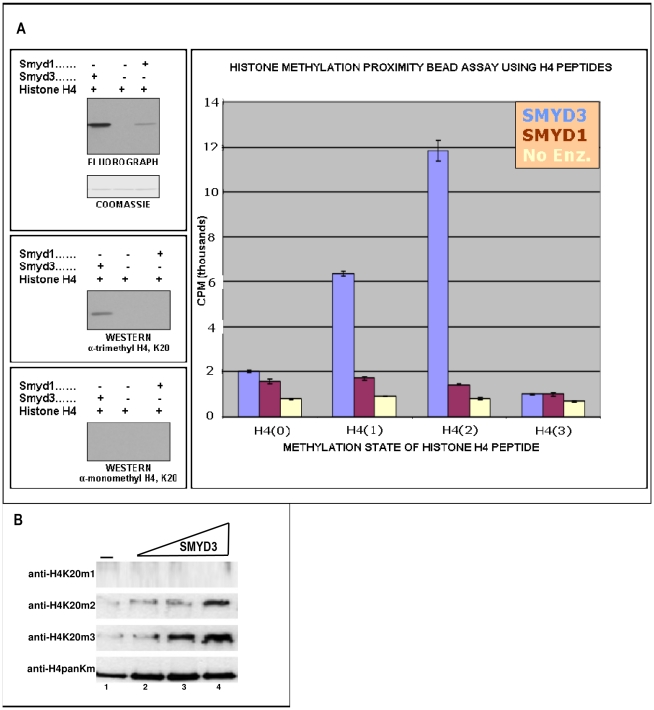
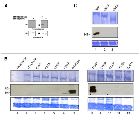
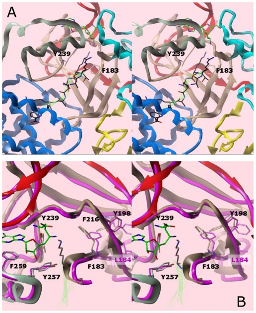
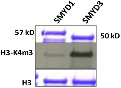
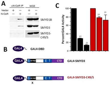
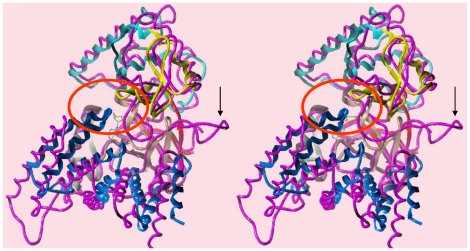
The SET and MYND Domain (SMYD) proteins comprise a unique family of multi-domain SET histone methyltransferases that are implicated in human cancer progression. Here we report an analysis of the crystal structure of the full length human SMYD3 in a complex with an analog of the S-adenosyl methionine (SAM) methyl donor cofactor. The structure revealed an overall compact architecture in which the "split-SET" domain adopts a canonical SET domain fold and closely assembles with a Zn-binding MYND domain and a C-terminal superhelical 9 α-helical bundle similar to that observed for the mouse SMYD1 structure. Together, these structurally interlocked domains impose a highly confined binding pocket for histone substrates, suggesting a regulated mechanism for its enzymatic activity. Our mutational and biochemical analyses confirm regulatory roles of the unique structural elements both inside and outside the core SET domain and establish a previously undetected preference for trimethylation of H4K20.


Conflict of interest statement

Competing Interests: KF, AC, LA, and SS were employed by OSI Pharmaceuticals at the time of the contribution and are currently employed at Coferon, Inc.; employed at OSI Pharmaceuticals, a wholly owned subsidiary of Astellas Pharma; self-employed (DiscoverElucidations); and employed at Abbott, respectively. FP and SE were employed at SGX Pharmaceuticals at the time of the contribution and are currently employed at AltheaDx and Eli Lilly, respectively. This does not alter the authors' adherence to all the PLoS ONE policies on sharing data and materials.

Figures

Figure 1
Figure 1. Structure of SMYD3 and its paralogs.
(A) Linear representation of domain structures of SMYDs1–5. The split SET domain is shown in red (N-SET) and tan (C-SET); the MYND domain is represented in yellow and the cysteine-rich post-SET domain is displayed in pale green. Starting and ending amino acids are indicated. (B) Ribbon representation of the structure of SMYD3-Sinefungin at 1.85Å resolution in cross-eye stereo. The SET domain of SMYD3 is split into the N-SET (red) and the C-SET (tan) by an intervening MYND domain (yellow) and a Rubisco-LSMT-like I-SET region (cyan). The post-SET motif (pale green) precedes a long (∼150 residue) C-terminal domain (CTD, blue). Positions of Sinefungin (green carbons) and zinc atoms (spheres) are indicated.
Figure 2
Figure 2. SMYD3 preferentially catalyzes histone 4 lysine methylation in vitro.
(A, B) SMYD3 purified from baculovirus methylates all histones (H4>>H2A>H3>H2B) in vitro. Histone methyltransferase (HMTase) assays employed mixed histones from HeLa cells as substrate. Upper panel, fluorography showing 3H-incorporation into H3 (17 kD) and into species smaller bands (H2A/H2B and H4). Lower panel, Coomassie-stained SDS-PAGE gel used to verify equal loading. (C) SMYD3 purified from bacteria methylates histones H4>>H3>H2A in vitro. Recombinant histones or mixed histones were used, as indicated, for substrate. Fluorography is shown and the bands corresponding to each histone are indicated.
Figure 3
Figure 3. SMYD3 trimethylates H4-K20 preferentially.
(A) SMYD3 trimethylates H4-K20. Right panel: unmethylated [H4(0)], mono-[H4(1)], di-[H4(2)], and, as a negative control, tri-methylated [H4(3)] peptides were employed in an in vitro HMTase proximity bead assay with baculoviral SMYD3 and SMYD1 (negative control). Degree of methylation was measured by scintillation counting in CPM. Left panels: Western analysis using anti-mono- and trimethyl-specific antibodies (Upstate) confirm in vitro specificity of SMYD3 for H4-K20me3. (B) SMYD3 preferentially trimethylates H4-K20 in reconstituted chromatin. Recombinant oocyte nucelosomes were assembled into chromatin, followed by in vitro HMTase assays and SDS-PAGE resolution of reaction products. SMYD3 inputs were increased from 0.5 µg to 2.4 µg, (triangle above lanes), and western analyses were performed with the indicated histone H4 methylation state-specific antibodies (middle panels), with a pan-anti-H4 (lower panel) providing a loading control for chromatin input.
Figure 4
Figure 4. Mutational analysis of residues critical to SMYD3 structure and function.
(A) Wild-type SMYD3, but not catalytic point mutant Y239F, methylates recombinant H3 and H4 in an in vitro HMTase assay. Upper panels: Fluorographs with bands corresponding to H3 (left) and H4 (right) indicated. Lower panels: Coomassie-stained PVDF membranes used to verify equal loading. (B) Substitution and (C) truncation mutants, constructed in E coli as described in Methods, were compared in in vitro HMTase assays to wildtype SMYD3 and to SET7/9. Inputs (upper panels, ∼500 ng) were assayed for 3H-SAM incorporation (middle panels) either on recombinant histone 4 or mixed histones, as indicated (lower panels). Alanine substitutions of most SMYD3 residues predicted to be catalytically essential eliminate HMTase activities. An exception is T184/A which, as described in the text, appears to affect H3-H4 substrate specificity (note change in relative ratios of H3/H4). N-terminal truncation through position 44 removes the entire N-SET domain, while truncation through position 74 eliminates both the N-SET domain and half the MYND domain.
Figure 5
Figure 5. Model of the SMYD3-Sinefungin active site with the H3K4me1 peptide in cross-eye stereo.
The peptide, colored black, and with backbone traced in green ribbon, was taken from an overlay with the SET9 ternary structure. (A) Ribbon representation of the ternary complex. Substrate methyl donor and peptide are indicated as green wire bonds and ribbons, respectively. Domain colors for SMYD3 correspond to those in Fig. 1. The aromatic cage residues (Y239, F183) at the end of the lysine channel are shown explicitly. (B) Overlay of SMYD1 (magenta, PDB accession #3N71) and SMYD3 (colored by domain) proteins in the ternary model. Numbering of residues is for SMYD3.
Figure 6
Figure 6. SMYD3 is more efficient than SMYD1 in trimethylation of their common substrate, H3-K4.
In vitro assays were carried out using bacterially expressed/purified 6XHis-SMYDs and recombinant H3 (top and lower, respectively, Coomassie-stain panels). Center panel: Autoradiography of the anti-H3-K4me3 (UpState) western blot.
Figure 7
Figure 7. An intact SMYD3 MYND domain is required for association with N-CoR and for transcriptional repression.
(A) N-CoR co-immunoprecipitates with wildtype SMYD3 but not with SMYD3 MYND domain point mutant C49/S. 293T cells were co-transfected with N-CoR, N-terminal myc-tagged SMYD3 constructs indicated, and with empty vector (vector). 48 hours post-transfection, whole cell RIPA lysates (WCL) were prepared. Fractions of the lysates were subjected to anti-N-CoR co-immunoprecipitation and the remaining 50% served as input. Western analysis was performed with anti-myc antibodies. Myc-SMYD1B, previously shown to interact with N-CoR served as a positive control. (B) Schematic of GAL4-DNA binding domain (DBD) and GAL4-fusion constructs for wild type (GAL4-SMYD3) and MYND domain-mutated (GAL4-SMYD3-C49/S) two hybrid transcription assays. X denotes the location of the C49/S mutation. (C) GAL4-SMYD3 but not GAL4-SMYD3-C49/S represses transcription of a GAL4-UAS containing luciferase reporter. 293T cells were transiently co-transfected with the 5XGAL4-SV40-luciferase reporter (1 µg) together with GAL4-DBD, or with 1 or 2 µg (indicated as 1X or 2X) of GAL4-SMYD3 (black bars) or GAL4-SMYD3-C49/S (red bars). Transfection efficiencies were normalized to co-transfected renilla luciferase, and percent GAL4 activity was determined in relation to GAL4-DBD set at 100%.
Figure 8
Figure 8. Unique carboxyl terminal domain (CTD).
Comparison of the CTD orientations in SMYD1 (magenta) and SMYD3 in cross-eye stereo. The larger loop structure in SMYD1 (indicated by the arrow) forces the CTD assembly to shift such that the C-terminus no longer contacts the MYND domain, as in SMYD3. The contacts between the two domains lie within the red circle.

References

    1. Venters B, Pugh BF. Chromatin meets RNA polymerase II. Genome Biology. 2007;8:319. - PMC - PubMed
    1. Bird AP. CpG-rich islands and the function of DNA methylation. Nature. 1986;321:209–213. - PubMed
    1. Cheng B, Price DH. Properties of RNA polymerase II elongation complexes before and after the P-TEFb-mediated transition into productive elongation. J Biol Chem. 2007;282:21901–21912. - PubMed
    1. McCabe MT, Brandes JC, Vertino PM. Cancer DNA methylation: molecular mechanisms and clinical implications. Clin Cancer Res. 2009;15:3927–3937. - PMC - PubMed
    1. Hamamoto R, Furukawa Y, Morita M, Iimura Y, Silva FP, et al. SMYD3 encodes a histone methyltransferase involved in the proliferation of cancer cells. Nat Cell Biol. 2004;6:731–740. - PubMed

Publication types

LinkOut - more resources