. 2011 Jan;2(1):46-55.
doi: 10.1177/1947601911405045.

The Putative PAX8/PPARγ Fusion Oncoprotein Exhibits Partial Tumor Suppressor Activity through Up-Regulation of Micro-RNA-122 and Dominant-Negative PPARγ Activity

Affiliations

The Putative PAX8/PPARγ Fusion Oncoprotein Exhibits Partial Tumor Suppressor Activity through Up-Regulation of Micro-RNA-122 and Dominant-Negative PPARγ Activity

Honey V Reddi et al. Genes Cancer. 2011 Jan.

Abstract

In vitro studies have demonstrated that the PAX8/PPARγ fusion protein (PPFP), which occurs frequently in follicular thyroid carcinomas (FTC), exhibits oncogenic activity. However, paradoxically, a meta-analysis of extant tumor outcome studies indicates that 68% of FTC-expressing PPFP are minimally invasive compared to only 32% of those lacking PPFP (χ(2) = 6.86, P = 0.008), suggesting that PPFP favorably impacts FTC outcomes. In studies designed to distinguish benign thyroid neoplasms from thyroid carcinomas, the previously identified tumor suppressor miR-122, a major liver micro-RNA (miR) that is decreased in hepatocellular carcinoma, was increased 8.9-fold (P < 0.05) in all FTC versus normal, 9.2-fold in FTC versus FA (P < 0.05), and 16.8-fold (P < 0.001) in FTC + PPFP versus FTC - PPFP. Constitutive expression of PPFP in the FTC-derived cell line WRO (WRO-PPFP) caused a 5-fold increase of miR-122 expression (P < 0.05) and a striking 5.1-fold reduction (P < 0.0001) in tumor progression compared to WRO-vector cells in a mouse xenograft model. Constitutive expression of either miR-122 or a dominant-negative PPARγ mutant in WRO cells was less effective than PPFP at inhibiting xenograft tumor progression (1.8-fold [P < 0.001] and 1.7-fold [P < 0.03], respectively). PPFP-induced up-regulation of miR-122 expression was independent of its known dominant-negative PPARγ activity. Up-regulation of miR-122 negatively regulates ADAM-17, a known downstream target, in thyroid cells, suggesting an antiangiogenic mechanism in thyroid carcinoma. This latter inference is directly supported by reduced CD-31 expression in WRO xenografts expressing PPFP, miR-122, and DN-PPARγ. We conclude that, in addition to its apparent oncogenic potential in vitro, PPFP exhibits paradoxical tumor suppressor activity in vivo, mediated by multiple mechanisms including up-regulation of miR-122 and dominant-negative inhibition of PPARγ activity.

Keywords: PAX8/PPARγ; follicular thyroid carcinoma; fusion protein; miR-122; tumor suppressor.


Conflict of interest statement

The author(s) declared no potential conflicts of interest with respect to the authorship and/or publication of this article.

Figures

Figure 1.
Figure 1.
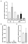
PPFP up-regulates the expression of miR-122 in FTC. Levels of miR-122 were evaluated using qRT-PCR. (A) Twelve fresh-frozen FTC, 6 containing PPFP and 6 without PPFP, (B) 10 formalin-fixed, paraffin-embedded FTC, 4 with PPFP and 6 without PPFP, (C) 7 normal thyroid tissue samples versus 14 FA, and (D) a comparison of miR-122 levels in NT-vector and NT-PPFP cells in vitro versus tumor xenografts in vivo. Asterisks indicate statistical significance at the P < 0.05 level. Error bars indicate SEM.
Figure 2.
Figure 2.
Overexpression of miR-122 in FTC-derived WRO cells inhibits tumor progression in nude mouse xenografts. (A) Stable transfection of a miR-122 precursor in WRO cells resulted in an approximate 8,000-fold increase (P < 0.005) in miR-122 expression as evaluated by quantitative RT-PCR. Expression of miR-122 in WRO cells results in a modest but significant (11%) increase in cell growth (B) and a significant inhibition (~2-fold) of tumor progression as assessed by repeated-measures (RM) ANOVA (P = 0.0082 and 0.0011, respectively). The values in B represent mean ± SD from 3 independent experiments, each done in triplicate, and values in C represent mean ± SEM from 16 and 10 independent tumors each for WRO-miR–null and WRO–miR-122 cells, respectively. Asterisks indicate statistical significance at the P < 0.05 level.
Figure 3.
Figure 3.
Stable expression of PPFP in FTC-derived WRO cells up-regulates miR-122 expression and inhibits xenograft tumor progression. The FTC-derived cell line WRO was genetically engineered to stably express PPFP using the vector shown in A. RT-PCR of WRO-PPFP cells (B) verifies expression of PPFP. (C) Expression of PPFP resulted in a 19% (P < 0.0001) inhibition of cell growth in vitro. (D) Subcutaneous injection of 5 × 10⁶ cells of WRO-PPFP cells into the right and left flanks of athymic nude mice was monitored over a period of 6 weeks and tumor volume measured and plotted as a function of time. Expression of PPFP in WRO cells results in significant inhibition (5.3-fold) of tumor progression as assessed by repeated-measures (RM) ANOVA (P = 0.0001). (E) Quantitation of miR-122 expression by RT-PCR demonstrated a significant increase (P < 0.05) in the WRO-PPFP cells and xenograft tumors versus the vector control. The values in C represent mean ± SD from 3 independent experiments, each done in triplicate. Values in D and E represent mean ± SEM from 8 independent tumors each for WRO-vector and WRO-PPFP cells. Asterisks indicate statistical significance at the P < 0.05 level.
Figure 4.
Figure 4.
PPFP functions in part by dominant-negative inhibition of PPARγ. (A) Scheme for generating the DN-PPARγ expression construct. (B) Expression of PPARγ (top panel), DN-PPARγ (middle panel), and β-actin (bottom panel) proteins as demonstrated by Western blotting of WRO-vector, -PPFP, and -DN-PPARγ cell extracts. (C) WRO-vector, -PPFP, or -DN-PPARγ cells were transiently transfected with the PPRE-Luc reporter and assayed for PPARγ activity, which is expressed as a percentage of WRO-vector–transfected cells. (D) The DN-PPARγ mutant resulted in a 29% (P < 0.0001) inhibition of cell growth in vitro. (E) Subcutaneous injection of 5 × 10⁶ cells of WRO-vector, -PPFP, or -DN-PPARγ cells into the right and left flanks of athymic nude mice was monitored over a period of 6 weeks and tumor volume measured. Expression of DN-PPARγ in WRO cells results in significant inhibition (1.8-fold) of tumor progression as assessed by repeated-measures (RM) ANOVA (P = 0.0074) but was not as effective as PPFP. miR-122 expression was not up-regulated when PPARγ activity was inhibited either with a DN-PPARγ mutant (F) or when WRO-vector cells were treated with GW9662, a potent PPARγ antagonist (H), which significantly inhibited PPARγ (PPRE) activity (G). Data in C, D, and F through H represent a mean of 3 experiments performed in triplicate. Data in E represent a mean of 8, 11, and 16 tumors each for vector, PPFP, and DN-PPARγ cells, respectively. Asterisks indicate statistical significance at the P < 0.05 level. Error bars indicate SEM.
Figure 5.
Figure 5.
ADAM-17 is down-regulated in the presence of miR-122. Quantitation of ADAM-17 transcript demonstrates a significant down-regulation in the presence of miR-122, which is restricted to the WRO–miR-122 (P < 0.0005) and WRO-PPFP (P < 0.005) cells (A) and FTC tumors containing PPFP (B). Normal thyroid tissues and follicular adenomas (FA) do not show reduction in ADAM-17 expression (C). Asterisks indicate statistical significance at the P < 0.05 level.
Figure 6.
Figure 6.
Neovascularization of WRO xenografts is inhibited by PPFP, DN-PPARγ, and miR-122 expression. (A) Immunohistochemical staining of WRO-vector, -PPFP, -DN-PPARγ, and –miR-122 xenograft tumors by CD-31 for quantitation of microvessel density (B) demonstrated that neovascularization is decreased 2.1-, 1.7-, and 3.4-fold in PPFP, miR-122, and DN-PPARγ xenograft tumors, respectively (P < 0.05), compared to WRO-vector xenografts. Asterisks indicate statistical significance at the P < 0.05 level. Error bars indicate SEM.

References

    1. Farahati J, Geling M, Mader U, et al. Changing trends of incidence and prognosis of thyroid carcinoma in lower Franconia, Germany, from 1981-1995. Thyroid. 2004;14:141-7 - PubMed
    1. Eberhardt NL, Grebe SK, McIver B, Reddi HV. The role of the PAX8/PPARgamma fusion oncogene in the pathogenesis of follicular thyroid cancer. Mol Cell Endocrinol. 2010;321:50-6 - PMC - PubMed
    1. Nambiar M, Kari V, Raghavan SC. Chromosomal translocations in cancer. Biochim Biophys Acta. 2008;1786:139-52 - PubMed
    1. Gregory Powell J, Wang X, Allard BL, et al. The PAX8/PPARgamma fusion oncoprotein transforms immortalized human thyrocytes through a mechanism probably involving wild-type PPARgamma inhibition. Oncogene. 2004;23:3634-41 - PubMed
    1. Au AY, McBride C, Wilhelm KG, Jr., et al. PAX8-peroxisome proliferator-activated receptor gamma (PPARgamma) disrupts normal PAX8 or PPARgamma transcriptional function and stimulates follicular thyroid cell growth. Endocrinology. 2006;147:367-76 - PubMed

LinkOut - more resources