. 2011 Apr;2(4):453-65.
doi: 10.1177/1947601911408893.

p53 Isoforms: An Intracellular Microprocessor?

Affiliations

p53 Isoforms: An Intracellular Microprocessor?

Marie P Khoury et al. Genes Cancer. 2011 Apr.

Abstract

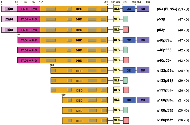
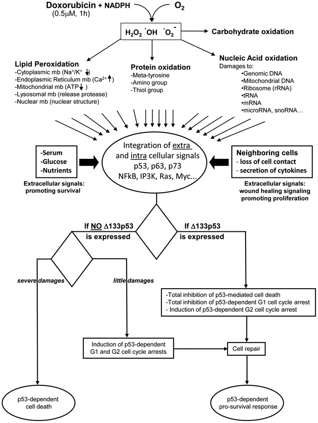
Normal function of the p53 pathway is ubiquitously lost in cancers either through mutation or inactivating interaction with viral or cellular proteins. However, it is difficult in clinical studies to link p53 mutation status to cancer treatment and clinical outcome, suggesting that the p53 pathway is not fully understood. We have recently reported that the human p53 gene expresses not only 1 but 12 different p53 proteins (isoforms) due to alternative splicing, alternative initiation of translation, and alternative promoter usage. p53 isoform proteins thus contain distinct protein domains. They are expressed in normal human tissues but are abnormally expressed in a wide range of cancer types. We have recently reported that p53 isoform expression is associated with breast cancer prognosis, suggesting that they play a role in carcinogenesis. Indeed, the cellular response to damages can be switched from cell cycle arrest to apoptosis by only manipulating p53 isoform expression. This may provide an explanation to the hitherto inconsistent relationship between p53 mutation, treatment response, and outcome in breast cancer. However, the molecular mechanism is still unknown. Recent reports suggest that it involves modulation of gene expression in a p53-dependent and -independent manner. In this review, we summarize our current knowledge about the biological activities of p53 isoforms and propose a molecular mechanism conciliating our current knowledge on p53 and integrating p63 and p73 isoforms in the p53 pathway.

Keywords: apoptosis; cell cycle; promoter; splice; tumor.


Conflict of interest statement

The author(s) declared no potential conflicts of interest with respect to the research, authorship, and/or publication of this article.

Figures

Figure 1.
Figure 1.
The human p53 gene expresses 12 distinct p53 protein isoforms. Schematic representation of the domains of human p53 isoform proteins including the 2 transactivation domains (TADI [light purple] and TADII [dark purple]), the DNA-binding domain (orange), the C-terminal domain comprised of the nuclear localization signal (NLS [yellow]), the oligomerization domain (OD [blue]), and the basic region (BR [violet]). The gray boxes represent the 5 highly conserved regions defining the p53 protein family. The amino acid positions defining the different p53 domains are indicated. The C-terminal domains of p53β (DQTSFQKENC) and p53γ (MLLDLRWCYFLINSS) are indicated with a green and pink box, respectively. The molecular weight of each p53 isoform protein is indicated.
Figure 2.
Figure 2.
p53AS: protein homology with p53β and biochemical activities. (A) Protein homology between human p53b ((h)p53b) and murine p53AS ((m)p53AS) isoforms. Conserved amino acids are highlighted in red. (B) Biochemical properties of mouse FLp53 and p53AS. (a) The intracellular localization, DNA binding, oligomerization, and (b) gene transactivation capacities (determined by luciferase assay) are highlighted. ND = not done; + = positive effect; ++ = stronger effect. Information obtained from Arai et al., Kulesz-Martin et al., Wu et al., Miner and Kulesz-Martin, Wu et al., Almog et al., Wolkowicz et al., Almog et al., Almog et al., and Huang et al..
Figure 3.
Figure 3.
Total number of p53RE sequences that can be written RRRCWWGYYY with 0 to 3 errors.
Figure 4.
Figure 4.
Extracellular and intracellular signals integrated by a single cell in response to a low dose of doxorubicin. Oxidation of doxorubicin generates free radicals that react with lipids, carbohydrates, proteins, and nucleic acids. Hence, doxorubicin causes lipid peroxidation, thus releasing the toxic content from the different subcellular organelles and causing loss of ATP production by mitochondria. Doxorubicin also causes loss of enzymatic activity and protein aggregation through thiol group oxidation, amino group oxidation, and formation of metatyrosine. Doxorubicin inhibits topoisomerase II activity, inducing DNA double-strand breaks. Moreover, doxorubicin binds covalently to DNA, inhibiting transcription and replication. Free radicals break all nucleic acids including mitochondrial DNA, ribosomal RNA, tRNA, mRNA, and microRNA. The damages are related to the concentration and duration of incubation with doxorubicin. The damaged cell is also in contact with surrounding cells, which secrete cytokines in response to doxorubicin treatment. If the neighboring cells die, the damaged cell loses its contact with the neighboring cells, triggering wound healing signaling. This is reinforced by the presence of nutrients, glucose, and growth factors. The extracellular and intracellular signals are integrated by cellular proteins (including p53, p63, and p73 isoforms), which will induce, in relation to the damages and the cell type, either prosurvival pathways (cell cycle arrest followed by cell repair, leading to proliferation or senescence) or cell death. The cell fate outcome is different if Δ133p53α is expressed or not. When Δ133p53α is expressed, p53-mediated apoptosis and G1 arrest are inhibited, while the p53-mediated G2 cell cycle arrest is promoted, allowing cell repair.

References

    1. El-Deiry WS, Kern SE, Pietenpol JA, Kinzler KW, Vogelstein B. Definition of a consensus binding site for p53. Nat Genet. 1992;1:45-9 - PubMed
    1. Bourdon JC, Deguin-Chambon V, Lelong JC, et al. Further characterisation of the p53 responsive element: identification of new candidate genes for trans-activation by p53. Oncogene. 1997;14:85-94 - PubMed
    1. Lane D, Levine A. p53 research: the past thirty years and the next thirty years. Cold Spring Harb Perspect Biol. 2010;2:a000893 - PMC - PubMed
    1. Bertheau P, Espie M, Turpin E, et al. TP53 status and response to chemotherapy in breast cancer. Pathobiology. 2008;75:132-9 - PubMed
    1. Murray-Zmijewski F, Slee EA, Lu X. A complex barcode underlies the heterogeneous response of p53 to stress. Nat Rev Mol Cell Biol. 2008;9:702-12 - PubMed

LinkOut - more resources