. 2011 Jun;5(3):185-91.
doi: 10.4162/nrp.2011.5.3.185. Epub 2011 Jun 21.

Chestnut extract induces apoptosis in AGS human gastric cancer cells

Affiliations

Chestnut extract induces apoptosis in AGS human gastric cancer cells

Hyun Sook Lee et al. Nutr Res Pract. 2011 Jun.

Abstract

In Korea, chestnut production is increasing each year, but consumption is far below production. We investigated the effect of chestnut extracts on antioxidant activity and anticancer effects. Ethanol extracts of raw chestnut (RCE) or chestnut powder (CPE) had dose-dependent superoxide scavenging activity. Viable numbers of MDA-MD-231 human breast cancer cells, DU145 human prostate cancer cells, and AGS human gastric cancer cells decreased by 18, 31, and 69%, respectively, following treatment with 200 µg/mL CPE for 24 hr. CPE at various concentrations (0-200 µg/mL) markedly decreased AGS cell viability and increased apoptotic cell death dose and time dependently. CPE increased the levels of cleaved caspase-8, -7, -3, and poly (ADP-ribose) polymerase in a dose-dependent manner but not cleaved caspase-9. CPE exerted no effects on Bcl-2 and Bax levels. The level of X-linked inhibitor of apoptosis protein decreased within a narrow range following CPE treatment. The levels of Trail, DR4, and Fas-L increased dose-dependently in CPE-treated AGS cells. These results show that CPE decreases growth and induces apoptosis in AGS gastric cancer cells and that activation of the death receptor pathway contributes to CPE-induced apoptosis in AGS cells. In conclusion, CPE had more of an effect on gastric cancer cells than breast or prostate cancer cells, suggesting that chestnuts would have a positive effect against gastric cancer.

Keywords: Chestnut; anticancer effect; antioxidant activity; ethanol extracts of chestnut; gastric cancer.


Figures

Fig. 1
Fig. 1
Effect of chestnut extracts on the viable cell numbers of MDA-MD-231, DU145, and AGS cells. (A) MDA-MB-231, (B) DU145, and (C) AGS cells were plated at a density of 50,000 cells/well in 24-well plates with DMEM/F12 supplemented with 10% FBS. One day after plating, the monolayers were serum-deprived with DMEM/F12 supplemented with 1% FBS serum-deprivation medium for 24 h. After serum deprivation, the cells were incubated in serum-deprivation medium in the absence or presence of 200 µg/mL viscous, aqueous extract of raw chestnut (RCW1), non-viscous, aqueous extract of raw chestnut (RCW2), aqueous extract of chestnut powder (CPW), ethanol extract of raw chestnut (RCE), or an ethanol extract of chestnut powder (CPE) for 24 hr. Cell numbers were estimated by the MTT assay. Each bar represents the mean ± SEM (n = 6). Means at a time without a common letter differ significantly, P < 0.05.
Fig. 2
Fig. 2
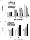
Effect of an ethanol extract of chestnut powder (CPE) on AGS cell viability and apoptosis. AGS cells were plated at a density of 50,000 cells/well in 24-well plates with DMEM/F12 supplemented with 10% FBS. One day after plating, the monolayers were serum-deprived with DMEM/F12 supplemented with 1% FBS serum-deprivation medium for 24 h. After serum deprivation, cells were incubated in serum-deprivation medium in the absence or presence of various concentrations of CPE. (A) Cell numbers were estimated by the MTT assay. (B) Apoptotic cells were detected with the Cell death detection ELISAPLUS. Each bar represents the mean ± SEM (n = 6). Means without a common letter differ significantly, P < 0.05.
Fig. 3
Fig. 3
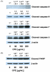
Effect of an ethanol extract of chestnut powder (CPE) on the levels of cleaved caspases (A) and poly (ADP-ribose) polymerase (PARP) (B) in AGS cells. Cells were plated and treated with various concentrations of CPE for 48 h as described in Fig. 2. Total cell lysates were analyzed by Western blotting with the indicated antibodies. Photographs of chemiluminescent detection of the blots, which are representative of three independent experiments, are shown. The relative abundance of each band to their own β-actin concentration was quantified and control levels were set to 1. The adjusted mean ± SEM is shown above each blot. Means without a common letter differ significantly, P < 0.05.
Fig. 4
Fig. 4
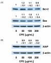
Effect of an ethanol extract of chestnut powder (CPE) on the levels of Bcl-2, Bax, and an inhibitor of apoptosis proteins (XIAP) in AGS cells. Cells were plated and treated with various concentrations of CPE for 48 h as described in Fig. 2. Total cell lysates were analyzed by Western blotting with the indicated antibodies. Photographs of chemiluminescent detection of the blots, which are representative of three independent experiments, are shown. The relative abundance of each band to their own β-actin concentration was quantified, and the control levels were set to 1. The adjusted mean ± SEM is shown above each blot. Means without a common letter differ significantly, P < 0.05.
Fig. 5
Fig. 5
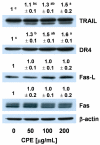
Effect of an ethanol extract of chestnut powder (CPE) on the levels of tumor necrosis factor-related apoptosis including ligand (TRAIL), DR4, Fas, and Fas-L in AGS cells. Cells were plated and treated with various concentrations of CPE for 48 h as described in Fig. 2. Total cell lysates were analyzed by Western blotting with the indicated antibodies. Photographs of chemiluminescent detection of the blots, which are representative of three independent experiments, are shown. The relative abundance of each band to their own β-actin concentration was quantified, and the control levels were set to 1. The adjusted mean ± SEM is shown above each blot. Means without a common letter differ significantly, P < 0.05.

Similar articles

Cited by

References

    1. Paillet FL. Chestnut: history and ecology of a transformed species. J Biogeogr. 2002;29:1517–1530.
    1. Kim JS, Jung BH, Joo RW, Choi SI. Marketing of chestnut and economic analysis of chestnut cultivation. Korean J For Econ. 2004;12:12–21.
    1. Heo J. Doguibogam. Seoul: Hanmibook press; 2001. p. 1153.
    1. Vekiari SA, Gordon MH, García-Macías P, Labrinea H. Extraction and determination of ellagic acid content in chestnut bark and fruit. Food Chem. 2008;110:1007–1011. - PubMed
    1. Barreira JCM, Ferreira ICFR, Oliveira MBPP, Pereira JA. Antioxidant activities of the extracts from chestnut flower, leaf, skins and fruit. Food Chem. 2008;107:1106–1113.