. 2011 Dec;60(12):1739-50.
doi: 10.1007/s00262-011-1069-4. Epub 2011 Jul 21.

A critical role for regulatory T cells in driving cytokine profiles of Th17 cells and their modulation of glioma microenvironment

Affiliations

A critical role for regulatory T cells in driving cytokine profiles of Th17 cells and their modulation of glioma microenvironment

Gabriele Cantini et al. Cancer Immunol Immunother. 2011 Dec.

Abstract

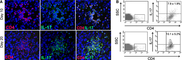
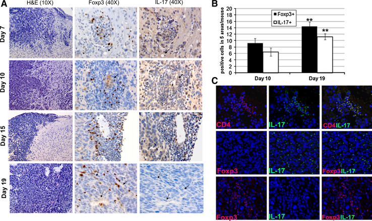
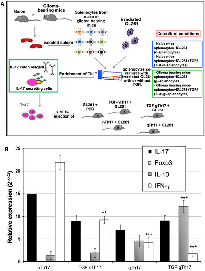
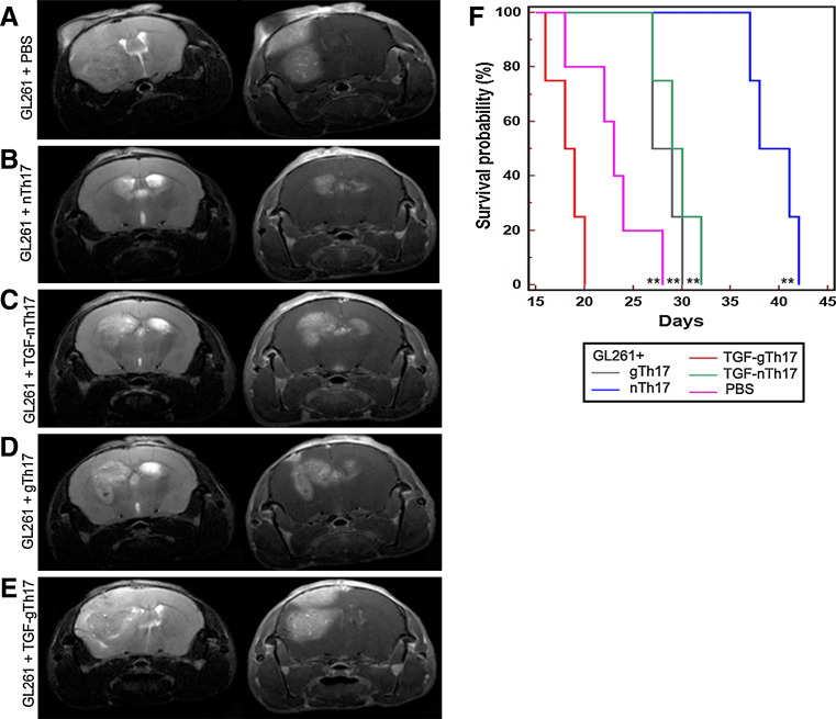
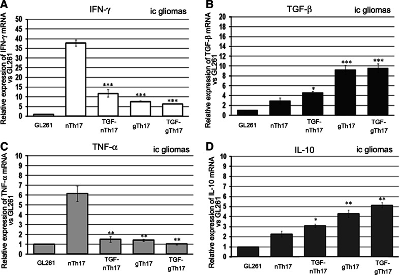
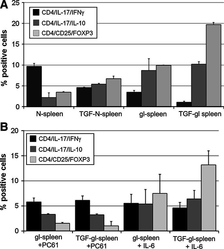
IL-17A, produced by Th17 cells, may play a dual role in antitumor immunity. Using the GL261-glioma model, we investigated the effects of Th17 cells on tumor growth and microenvironment. Th17 cells infiltrate mouse gliomas, increase significantly in a time-dependent manner similarly to Treg and do not express Foxp3. To characterize the direct effects of Th17 cells on GL261 murine gliomas and on tumor microenvironment, we isolated IL-17-producing cells enriched from splenocytes derived from naïve (nTh17) or glioma-bearing mice (gTh17) and pre-stimulated in vitro with or without TGF-β. Spleen-derived Th17 cells co-expressing IL-17, IFN-γ and IL-10, but not Treg marker Foxp3, were co-injected intracranially with GL261 in immune-competent mice. Mice co-injected with GL261 and nTh17 survived significantly longer than gTh17 (P < 0.006) and gliomas expressed high level of IFN-γ and TNF-α, low levels of IL-10 and TGF-β. In vitro IL-17 per se did not exert effects on GL261 proliferation; in vivo gliomas grew equally well intracranially in IL-17 deficient and wild-type mice. We further analyzed relationship between Th17 cells and Treg. Treg were significantly higher in splenocytes from glioma-bearing than naïve mice (P = 0.01) and gTh17 produced more IL-10 than IFN-γ (P = 0.002). In vitro depletion of Treg using PC61 in splenocytes from glioma-bearing mice causes increased IL-17/IFN-γ cells (P = 0.007) and decreased IL-17/IL-10 cells (P = 0.03). These results suggest that Th17 polarization may be induced by Treg and that Th17 cells in gliomas modulate tumor growth depending on locally produced cytokines.


Conflict of interest statement

The authors declare that they have no competing interests.

Figures

Fig. 1
Fig. 1
Increment of Th17 cells infiltration during glioma progression. a On day 10, only few CD4+ cells are detected inside the tumor area: most of double-positive lymphocytes, recruited from peripheral lymphoid organs, are concentrated around the tumor vessels. On day 20, after GL261 injection, the microenvironment immunocharacterization shows an increment of CD4+ IL-17+ population and double-positive cells are distributed as cellular cluster within the tumor area. Isotype control cells are not detectable in GL261-glioma (data not shown). b Infiltrating Th17 cells investigated by flow cytometry increased from 7.9 ± 1.9 on day 10 to 13.1 ± 5.2 on day 20 (P = 0.03). The panels show a representative dot plot and mean percentage value ± SD obtained from three different evaluations at each time point
Fig. 2
Fig. 2
Treg and Th17 cells are differentially distributed into the tumor mass. a Arrows indicate the same blood vessels in adjacent sections of tumors evaluated for the presence of infiltrating Foxp3 and IL-17 positive cells at different time points; 7, 10, 15 and 19 days after tumor implantation. Two representative mice for each time point have been investigated, and representative images are displayed. b Quantitative analysis is summarized in histogram, results are expressed as mean ± SD of positive cells in 5 areas/mouse/time point (number of positive cells/5 different 40× HPF). c Immunofluorescence of GL261-glioma on day 19 showed infiltrating CD4+ IL-17+ population double-positive cells, but negative for Treg marker Foxp3. Foxp3-positive cells do not co-express IL-17
Fig. 3
Fig. 3
Spleen-derived Th17 cells express IL-17, IFN-γ and IL-10 and do not express Foxp3. a Schematic experimental procedure. Spleen from naïve C57BL6N and glioma-bearing mice was surgically removed, and splenocytes were co-cultured in vitro with irradiated GL261 tumor cells in the presence or absence of rhTGF-β1. IL-17 secreting cells from different co-culture conditions (see the box below) were isolated and enriched immunomagnetically. Th17 cells were co-injected intracranially (ic) and subcutaneously (sc) with GL261 cells in immune-competent mice. b Th17 cells enriched by immunomagnetic separation were characterized by RT-PCR. All fractions express high levels of IL-17 and different expression of IFN-γ and IL-10 (*P < 0.01; **P < 0.0001; ***P < 0.00005). As shown in histogram, Foxp3 has not been detected
Fig. 4
Fig. 4
Th17 cells play a role on GL261-glioma growth. a MRI performed 16 days after tumor implantation shows that lesion derived from GL261 + PBS was large. b nTh17 cells provide a significant delay on tumor formation. T2 (b left, 20 slices; thickness 0.8 mm, no gap; TR: 2,658 ms/TE: 54 ms/RARE Factor 8/FOV: 2.20 × 2.20 cm2; averages 6; image matrix 256 × 256; in plane resolution 0.086 mm; acquisition time 8 min 30 s) and T1 with contrast medium representative images show the total absence of mass lesion compared to the other groups of co-injection (b right 18 slices; thickness 0.8 mm, no gap; TR: 435 ms/TE: 11 ms/FOV: 2.20 × 2.20 cm2; averages 4; image matrix 256 × 256: in plane resolution 0.086 mm; acquisition time 7 min 26 s). T2 and T1 images of gliomas from GL261 plus gTh17 (d) showed expanded and lesions compared to nTh17 or TGF-nTh17 (c). Lesion from co-injection with TGF-gTh17 (e) is shown both in T1 as a homogeneous enhancing mass and in T2 images as a strong mass effect toward lateral ventricle. Images refer to one representative animal for each group (five mice for each group). f Kaplan–Meier analysis curves represent the survival time (in days) of five mice for each group of co-injection. Survival rate of mice injected with nTh17 cells was significantly higher than that of control mice (GL261 plus PBS P = 0.005) and of mice injected with TGF-gTh17 cells (P = 0.0067). Mice injected with TGF-gTh17 cells survived significantly lower than controls (P = 0.02)
Fig. 5
Fig. 5
Th17 cells modulate tumor microenvironment. RT-PCR analysis of different gene expression performed on paraffin-embedded ic-gliomas. mRNA levels in each group were normalized to the relative quantity of b2m; relative expression of different cytokines was evaluated versus the GL261 plus PBS. a and c nTh17 tumors show an enhanced expression in IFN-γ and TNF-α compared to GL261 and TGF-gTh17 tumors. On the contrary, TGF-β and IL-10 (b and d) are strongly increased in gTh17-treated mice (P value: *P = 0.01; **P < 0.001; ***P < 0.0001)
Fig. 6
Fig. 6
IL-17 has no effect on the GL261-glioma formation. a MRI 7 T images on a frontal plane. T2 (Left) and T1 after contrast medium (Right) images on day 8 after GL261 implantation performed on a wild-type (WT) and IL-17 KO (KO) mouse show a similar tumor engraftment. b Brain pictures of wt and IL-17 KO 12 days after tumor implantation (Upper) and Ki-67 staining (magnification 40×, Lower) show a similar proliferation ratio in wt and IL-17 KO mice. c MRI 7 T images on a frontal plane. T2 (Left) and T1 after contrast medium (Right) images 16 after GL261 implantation performed on a wild-type and IL-17 KO mouse. d Kaplan–Meier analysis curves represent the survival time (in days) of IL-17 KO compared to WT mice injected ic with GL261 cells. All mice died by day 29. The median survival of IL-17 KO was 27 days (n = 5, mean ± SD is 26.8 ± 2.2); the median of WT mice was 26.5 (n = 5, mean ± SD is 26.8 ± 1.7) (P = 0.9)
Fig. 7
Fig. 7
CD4+ CD25+ Foxp3+ cells (Treg) modulate the Th17 cytokine profile. a Intracellular FACS analysis of naïve and tumor bearing CD4+ lymphocytes. Treg are significantly higher in glioma-bearing mice versus naïve splenocytes (P = 0.01), and they increase significantly in the presence of TGF-β (P = 0.001 in gl-splenocytes; P = 0.02 in n-splenocytes). nTh17 produced higher levels of IFN-γ than IL-10 (P = 0.001) that appeared reduced by TGF-β (P = 0.01). On the contrary, gTh17 produced more IL-10 than IFN-γ (P = 0.002). The presence of TGF-β promoted a relevant decrease in IFN-γ (P = 0.01) and a significant increase in IL-10-producing T cells (P = 0.03). Notably, the absolute number of Th17 cells obtained from the same number of splenocytes did not change significantly (nTh17 8.1 × 105; TGF-nTh17 8 × 105; gTh17 7.9 × 105; TGF-gTh17 9.2 × 105). Data are relative of three independent experiments. b Intracellular FACS analysis of tumor bearing CD4+ lymphocytes. PC61 antibody (0.5 μg/1 × 106 cells) decreased Treg population from gl- and TGF-gl-splenocytes (P < 0.001), increasing IL-17/IFN-γ production (gTh17 P = 0.05; TGF-gTh17 P = 0.01) and decreasing IL-17/IL-10 secretion (gTh17 P = 0.0004; TGF-gTh17, P = 0.003). Data are statistically significant when they are compared to the same culture condition without PC61 (Fig. 7a). No significant differences (P > 0.05) were found in the same experiments performed with IL-6. In vitro data are represented as mean ± SD obtained combining percentage obtained from 24 to 48 h pre-stimulation of splenocytes

References

    1. Steinman L. A brief history of T(H)17, the first major revision in the T(H)1/T(H)2 hypothesis of T cell-mediated tissue damage. Nat Med. 2007;13:139–145. doi: 10.1038/nm1551. - DOI - PubMed
    1. Harrington LE, Hatton RD, Mangan PR, Turner H, Murphy TL, Murphy KM, Weaver CT. Interleukin 17-producing CD4+ effector T cells develop via a lineage distinct from the T helper type 1 and 2 lineages. Nat Immunol. 2005;6:1123–1132. doi: 10.1038/ni1254. - DOI - PubMed
    1. Stockinger B, Veldhoen M. Differentiation and function of Th17 T cells. Curr Opin Immunol. 2007;19:281–286. doi: 10.1016/j.coi.2007.04.005. - DOI - PubMed
    1. Das J, Ren G, Zhang L, Roberts AI, Zhao X, Bothwell AL, Van Kaer L, Shi Y, Das G. Transforming growth factor beta is dispensable for the molecular orchestration of Th17 cell differentiation. J Exp Med. 2009;206:2407–2416. doi: 10.1084/jem.20082286. - DOI - PMC - PubMed
    1. Laurence A, Tato CM, Davidson TS, Kanno Y, Chen Z, Yao Z, Blank RB, Meylan F, Siegel R, Hennighausen L, Shevach EM, O’shea JJ. Interleukin-2 signaling via STAT5 constrains T helper 17 cell generation. Immunity. 2007;26:371–381. doi: 10.1016/j.immuni.2007.02.009. - DOI - PubMed

Publication types