. 2011 Dec;14(4):443-55.
doi: 10.1007/s10456-011-9227-z. Epub 2011 Jul 21.

Anti-CXCL5 therapy ameliorates IL-17-induced arthritis by decreasing joint vascularization

Affiliations

Anti-CXCL5 therapy ameliorates IL-17-induced arthritis by decreasing joint vascularization

Sarah R Pickens et al. Angiogenesis. 2011 Dec.

Abstract

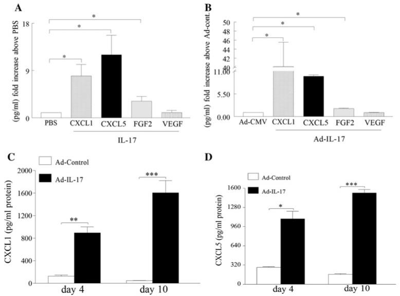
IL-17-induced joint inflammation is associated with increased angiogenesis. However, the mechanism by which IL-17 mediates angiogenesis is undefined. Therefore, the pathologic role of CXCL1 and CXCL5 was investigated in arthritis mediated by local expression of IL-17, employing a neutralizing antibody to each chemokine. Next, endothelial chemotaxis was utilized to examine whether endothelial migration was differentially mediated by CXCL1 and CXCL5. Our results demonstrate that IL-17-mediated disease activity was not affected by anti-CXCL1 treatment alone. In contrast, mice receiving anti-CXCL5 demonstrated significantly reduced clinical signs of arthritis, compared to the mice treated with IgG control. Consistently, while inflammation, synovial lining thickness, bone erosion and vascularization were markedly reduced in both the anti-CXCL5 and combination anti-CXCL1 and 5 treatment groups, mice receiving anti-CXCL1 antibody had clinical scores similar to the control group. In contrast to joint FGF2 and VEGF levels, TNF-α was significantly reduced in mice receiving anti-CXCL5 or combination of anti-CXCL1 and 5 therapies compared to the control group. We found that, like IL-17, CXCL1-induced endothelial migration is mediated through activation of PI3K. In contrast, activation of NF-κB pathway was essential for endothelial chemotaxis induced by CXCL5. Although CXCL1 and CXCL5 can differentially mediate endothelial trafficking, blockade of CXCR2 can inhibit endothelial chemotaxis mediated by either of these chemokines. These results suggest that blockade of CXCL5 can modulate IL-17-induced inflammation in part by reducing joint blood vessel formation through a non-overlapping IL-17 mechanism.


Figures

Fig. 1
Fig. 1
IL-17 induces production of CXCL1 in RA synovial fibroblasts and macrophages however, only in RA fibroblasts is CXCL1 expression synergistically induced by IL-17 and TNF-α. RA synovial tissue fibroblasts (a) and normal macrophages (d) were activated with IL-17 (50 ng/ml) for 0–8 h. Real-time RT-PCR was employed to identify CXCL1 (a and d) mRNA levels which were normalized to GAPDH. The results are presented as fold increase, compared with the 0 h time point (untreated cells). RA synovial tissue fibroblasts (c) and normal macrophages (e) were either untreated or incubated with DMSO or inhibitors to PI3K (LY294002; 10 μM), ERK (PD98059; 10 μM), JNK (SP600125; 10 μM) or p38 (SB203580; 10 μM) for 1 h. Thereafter cells treated with DMSO or inhibitors were subsequently activated with IL-17 (50 ng/ml) for 24 h and the media was collected from all conditions in order to quantify the levels of CXCL1 employing ELISA. b RA synovial tissue fibroblasts were either unstimulated or stimulated with IL-17 (50 ng/ml), TNF-α (10 ng/ml), or IL-17 plus TNF-α for 6 h. Cells were harvested, and CXCL1 mRNA levels were quantified by real-time RT-PCR which were normalized to GAPDH and presented as fold increase above PBS treatment (unstimulated cells). Values represent the mean ± SE. * Represents P < 0.05 and ** denotes P < 0.01, n = 3–5
Fig. 2
Fig. 2
In RA synovial fibroblasts and macrophages, IL-17 induces production of CXCL5 however only in RA fibroblasts is CXCL5 expression synergistically induced by IL-17 and TNF-α stimulation. RA synovial tissue fibroblasts (a) and normal macrophages (d) were activated with IL-17 (50 ng/ml) for 0–8 h. Real-time RT-PCR was employed to identify CXCL5 (a and d) mRNA levels which were normalized to GAPDH. The results are presented as fold increase, compared with the 0 h time point (untreated cells). RA synovial tissue fibroblasts (c) and normal macrophages (e) were either untreated or incubated with DMSO or inhibitors to PI3K (LY294002; 10 μM), ERK (PD98059; 10 μM), JNK (SP600125; 10 μM) or p38 (SB203580; 10 μM) for 1 h. Thereafter cells treated with DMSO or inhibitors were subsequently activated with IL-17 (50 ng/ml) for 24 h and the supernatants were collected from all conditions in order to quantify the levels of CXCL5 by ELISA. b RA synovial tissue fibroblasts were either unstimulated or stimulated with IL-17 (50 ng/ml), TNF-α (10 ng/ml), or IL-17 plus TNF-α for 6 h. Cells were harvested and CXCL5 mRNA levels were quantified by real-time RT-PCR which were normalized to GAPDH and presented as fold increase above PBS treatment (unstimulated cells). Values represent the mean ± SE. * Represents P < 0.05 and ** denotes P < 0.01, n = 3–5
Fig. 3
Fig. 3
IL-17 increases the expression of CXCL1 and CXCL5 in RA synovial tissue explants and experimental arthritis model. a RA synovial tissue explants were treated with PBS or IL-17 (100 ng/ml), tissues were harvested after 24 h and levels of CXCL1, CXCL5, FGF2 and VEGF were quantified by ELISA and normalized to PBS values. b Ad-IL-17 or Ad-CMV control (107 PFU) was injected intra-articularly into C57/BL6 mice. Ankles were harvested on day 10 and levels of CXCL1, CXCL5, FGF2 and VEGF were measured by ELISA and normalized to Ad-CMV. c, d Ankles from Ad-IL-17 and Ad-CMV treatment were harvested on days 4 and 10, and CXCL1 (c) and CXCL5 (d) levels were quantified by ELISA. Values are reported as mean ± SE. * Denotes P < 0.05, ** denotes P < 0.01 and *** denotes P < 0.005, n = 8–10
Fig. 4
Fig. 4
Neutralization of CXCL5 ameliorates IL-17-induced joint inflammation, synovial lining thickness, bone erosion and joint TNF-α levels. C57BL/6 mice were treated intraperitoneally with 30 μg (total of 210 μg was utilized over the course of treatment) IgG, anti-CXCL1, anti-CXCL5 or both anti-CXCL1 and 5 antibodies (Leinco Technologies) on days −4, −2, 0, 3, 5, 7 and 9 post-Ad injection. On day 0, Ad-IL-17 (107 PFU) was injected intra-articularly into the mouse ankle joint, and the joint circumference (a) was measured on days 0, 3, 5, 7 and 10 post-Ad-IL-17 injection and each experimental group consisted of 10–12 mice. b, c Inflammation, synovial lining and bone erosion (based on a 0–5 score) were determined using H&Estained sections by a blinded observer, n = 10 ankles. Changes in the levels of joint TNF-α (d) were measured in ankle homogenates obtained from different treatment groups by ELISA and were normalized by protein concentration, n = 7–9 ankles. Values demonstrate mean ± SE. * Denotes P < 0.05
Fig. 5
Fig. 5
Levels of joint FGF2 and VEGF were unaffected by anti-CXCL5 and combination therapy and anti-CXCL5 therapy downregulates IL-17-induced joint vascularization. Changes in the levels of joint FGF2 (a) and VEGF (b) were measured in ankle homogenates obtained from different treatment groups by ELISA and the data is presented as fold increase above the control IgG treatment, n = 7–9 ankles. IL-17-induced arthritis ankles treated with IgG, anti-CXCL1, anti-CXCL5 or the combination therapy were harvested on day 11 and were immunostained with Von willebrand factor (endothelial marker) (c) (original magnification × 200). Quantification of endothelial (d) staining from IL-17-induced arthritis ankles harvested on day 11, n = 8–9 ankles. Values demonstrate mean ± SE. * Denotes P < 0.05
Fig. 6
Fig. 6
Anti-CXCL5 treatment did not affect the circulating number of leukocytes, neutrophils and monocytes in IL-17-mediated arthritis model and CXCL1 and CXCL5 mediate endothelial migration through CXCR2 ligation. a On day 11 blood was collected by cardiac puncture of IL-17-induced arthritis ankles treated with IgG, anti-CXCL1, anti-CXCL5 or the combination therapy to measure blood cell count using a HemaVet 850 complete blood counter. Values are shown in thousands of cells per microliter of blood (k/μl, n = 10–12 mice). b HMVECs incubated with antibody to CXCR2 (10 μg/ml, R&D systems) were kept at 37°C for 2 h while cells were attaching to the membrane and chemotaxis was examined in response to CXCL1 and CXCL5 (1 and 20 ng/ml; for 2 h at 37°C), n = 2. Values represent fold increase chemotaxis above cells migrating in response to PBS shown as mean ± SE of two experiments in triplicate. * Represents P < 0.05
Fig. 7
Fig. 7
CXCL1 and CXCL5 induce endothelial migration through activating different signaling pathways. In order to determine the mechanism by which (a) CXCL1 and (c) CXCL5 activate HMVECs, cells were stimulated with these chemokines (20 ng/ml) for 0–65 min, and the cell lysates were probed for IκB,p-p38, p-AKT,and pERK and/or equal loading controls. To determine signaling pathways associated with b CXCL1 and d CXCL5-induced HMVEC migration, cells were treated with DMSO or inhibitors to NF-κB (MG-132; 1 and 5 μM), p38 (SB203580; 1 and 5 μM), PI3K (LY294002; 1 and 5 μM) and ERK (PD98059; 1 and 5 μM) 2 h in the Boyden chamber, n = 2. Values represent fold increase chemotaxis above cells migrating in response to PBS shown as mean ± SE of two experiments in triplicate. * Represents P< 0.05

References

    1. Firestein GS. Evolving concepts of rheumatoid arthritis. Nature. 2003;423:356–361. - PubMed
    1. Lubberts E, Koenders MI, Oppers-Walgreen B, et al. Treatment with a neutralizing anti-murine interleukin-17 antibody after the onset of collagen-induced arthritis reduces joint inflammation, cartilage destruction, and bone erosion. Arthritis Rheum. 2004;50:650–659. - PubMed
    1. Koenders MI, Lubberts E, van de Loo FA, et al. Interleukin-17 acts independently of TNF-alpha under arthritic conditions. J Immunol. 2006;176:6262–6269. - PubMed
    1. Lubberts E, Joosten LA, Oppers B, et al. IL-1-independent role of IL-17 in synovial inflammation and joint destruction during collagen-induced arthritis. J Immunol. 2001;167:1004–1013. - PubMed
    1. Koenders MI, Lubberts E, Oppers-Walgreen B, et al. Induction of cartilage damage by overexpression of T cell interleukin-17A in experimental arthritis in mice deficient in interleukin-1. Arthritis Rheum. 2005;52:975–983. - PubMed

Publication types

MeSH terms