

MR properties of brown and white adipose tissues
- PMID: 21780237
- PMCID: PMC3146031
- DOI: 10.1002/jmri.22623
MR properties of brown and white adipose tissues
Abstract
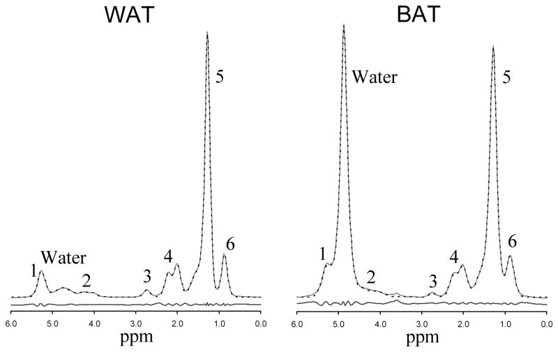
Purpose: To explore the MR signatures of brown adipose tissue (BAT) compared with white adipose tissue (WAT) using single-voxel MR spectroscopy.
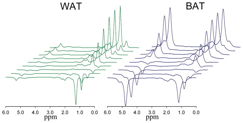
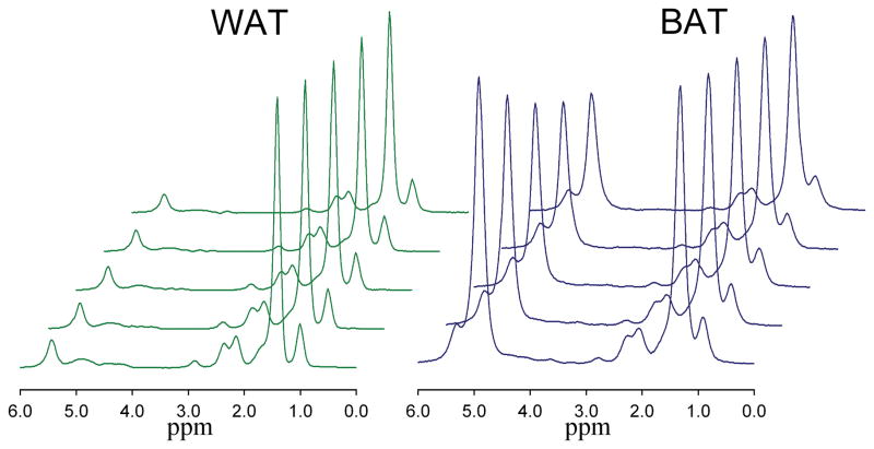
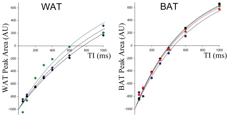
Materials and methods: (1) H MR STEAM spectra were acquired from a 3 Tesla clinical whole body scanner from seven excised murine adipose tissue samples of BAT (n=4) and WAT (n=3). Spectra were acquired at multiple echo times (TEs) and inversion times (TIs) to measure the T1, T2, and T2-corrected peak areas. A theoretical triglyceride model characterized the fat in terms of number of double bonds (ndb) and number of methylene-interrupted double bonds (nmidb).
Results: Negligible differences between WAT and BAT were seen in the T1 and T2 of fat and the T2 of water. However, the water fraction in BAT was higher (48.5%) compared with WAT (7.1%) and the T1 of water was lower in BAT (618 ms) compared with WAT (1053 ms). The fat spectrum also differed, indicating lower levels of unsaturated triglycerides in BAT (ndb=2.7, nmidb=0.7) compared with WAT (ndb=3.3, nmidb=1.0).
Conclusion: We have demonstrated that there are several key MR-based signatures of BAT and WAT that may allow differentiation on MR imaging.
Copyright © 2011 Wiley-Liss, Inc.
Figures
References
-
- Himms-Hagen J. Thermogenesis in brown adipose tissue as an energy buffer. Implications for obesity. N Engl J Med. 1984;311:1549–1558. - PubMed
-
- Cannon B, Nedergaard J. Brown adipose tissue: function and physiological significance. Physiol Rev. 2004;84:277–359. - PubMed
-
- Nedergaard J, Cannon B. The changed metabolic world with human brown adipose tissue: therapeutic visions. Cell Metab. 2010;11:268–272. - PubMed
-
- Enerback S. Human brown adipose tissue. Cell Metab. 2010;11:248–252. - PubMed
Publication types
MeSH terms
Substances
Grants and funding
LinkOut - more resources
Full Text Sources
Other Literature Sources
Medical
