. 2011 Sep;240(9):2127-41.
doi: 10.1002/dvdy.22702. Epub 2011 Jul 20.

Transforming growth factor Beta2 is required for valve remodeling during heart development

Affiliations

Transforming growth factor Beta2 is required for valve remodeling during heart development

Mohamad Azhar et al. Dev Dyn. 2011 Sep.

Abstract

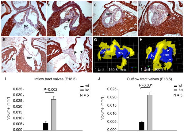
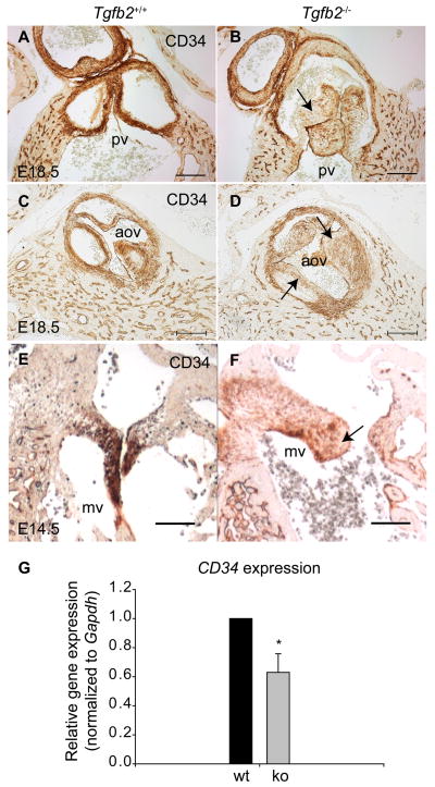
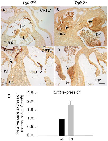
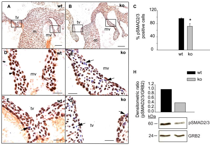
Although the function of transforming growth factor beta2 (TGFβ2) in epithelial mesenchymal transition (EMT) is well studied, its role in valve remodeling remains to be fully explored. Here, we used histological, morphometric, immunohistochemical and molecular approaches and showed that significant dysregulation of major extracellular matrix (ECM) components contributed to valve remodeling defects in Tgfb2(-/-) embryos. The data indicated that cushion mesenchymal cell differentiation was impaired in Tgfb2(-/-) embryos. Hyaluronan and cartilage link protein-1 (CRTL1) were increased in hyperplastic valves of Tgfb2(-/-) embryos, indicating increased expansion and diversification of cushion mesenchyme into the cartilage cell lineage during heart development. Finally, Western blot and immunohistochemistry analyses indicate that the activation of SMAD2/3 was decreased in Tgfb2(-/-) embryos during valve remodeling. Collectively, the data indicate that TGFβ2 promotes valve remodeling and differentiation by inducing matrix organization and suppressing cushion mesenchyme differentiation into cartilage cell lineage during heart development.


Figures

Fig. 1
Fig. 1
Cardiac valve thickening in Tgfb2-/- embryos. A-F: Immunohistochemistry showing cardiac valve morphology (HHF-35 staining for cardiac actin) in wild-type (A,C,E) and Tgfb2-/- (B,D,F) embryos at E18.5. Arrows denote thickened pulmonary (B), aortic (D) and mitral (F) valves in Tgfb2-/- embryos. G,H: Three-dimensional reconstructions of mitral valves of E18.5 wild-type and Tgfb2-/- embryos shown in (E and F) indicate leaflet thickening (arrow, H) and thickened chordae tendineae (arrowhead, H). Volume of the reconstructed mitral valves in Tgfb2-/- embryo (F,H) is 0.014 mm3 and 0.004 mm3 in wild-type embryo (E,G) All images (A-F) are representative of more than 9 wild-type/Tgfb2-/- embryo pairs. I,J: Morphometric comparison of inflow tract valves volume (I) and outflow tract valve volume between wild-type and Tgfb2-/- embryos at E18.5. Number of samples analyzed and the P values are given on the histograms. The total volume of inflow tract or outflow tract valves is significantly increased in Tgfb2-/- embryos as compared to wild-type embryos. Scale bar = 100 μm in (A-D), 200 μm (E,F). pv, pulmonary valves; aov, aortic valves; mv, mitral valves; m, myocardium; tv, tricuspid valves.
Fig. 2
Fig. 2
Dysregulated extracellular matrix organization in thickened valves of Tgfb2-/- embryos. A-D: Weigert's Resorcin-Fuchsin-staining of wild-type (A, B) and Tgfb2-/- (C, D) mitral valve leaflets at E18.5. Magnified views of regions indicated by the box in wild-type (A) and Tgfb2-/- (C) embryos are presented in (B) and (D), respectively, showing significantly decreased elastin fibers in Tgfb2-/- embryo compared to the wild-type embryo (D, arrow). E-F: Picrosirius Red-stained sections of wild-type (E) and Tgfb2-/- (F) embryos at E18.5 visualized in birefringent optics showing diffused collagen fibers in mitral valves of Tgfb2-/- embryos (arrows, F) as compared to wild-type embryo (E). G,H: Alcian blue staining showing excess accumulation of GAGs in inflow tract valves of Tgfb2-/- embryo (arrows in H) compared to the wild-type embryo (G) at E18.5. I,J: Immunohistochemistry indicating increased expression of HABP in mitral valves of Tgfb2-/- embryo (J) as compared to the wild-type embryo (I) at E18.5.K: Histogram showing the relative amount of hyaluronan in mitral valves as indicated by HABP florescence. The intensity of staining is graded semiquantitatively on a scale from 0 to 3, as described in details in Experimental Procedures section. The mean scores for mitral valves from more than three wild-type/Tgfb2-/- embryo pairs are presented. L,M: Real time PCR analyses on pooled cardiac tissue samples containing both outflow tract and inflow tract cushions of wild-type or Tgfb2-/- embryos at E14.5. Three biologically different pooled samples of wild-type and Tgfb2-/- embryos were assessed, as described in the Experimental Procedures. Each wild-type value is normalized to 1.0. The expression of Cspg2 and the hyaluronan receptor Rhamm is significantly upregulated in Tgfb2-/- embryos compared to wild-type embryos at E14.5 (*P= 0.0248 for Cspg2, *P= 2.7484e-3 for Rhamm) (L). The expression of both Fibrillin-1 (*P=0.0344) and Lox (*P=0.0154) is significantly reduced in Tgfb2-/- embryos as compared to wild-type embryos (M). All images (A-J) are representative of more than three wild-type/ Tgfb2-/- embryo pairs. Scale bar = 100 μm in (A, C), 20 μm (B, D), 50 μm in (E, F), 100 μm in (G,H), 50 μm in (I,J). wt, wild-type; ko, Tgfb2-/-; mv, mitral valves.
Fig. 3
Fig. 3
Impaired cushion cell differentiation in Tgfb2-/- embryos. A-D: Cardiac morphology (IHC: HHF-35, counterstained with hematoxylin) showing pulmonary valves in wild-type (A) and Tgfb2-/- (C) embryos and anti-BrdU-staining of their corresponding sections in wild-type (B) and Tgfb2-/- (D) embryos at E16.5. In (B,D), arrows indicate nuclear BrdU incorporating cells in pulmonary valves of developing hearts. All images (A-D) are representative of more than three wild-type/Tgfb2-/- embryo pairs. E: Morphometric comparison of cell density (# of cells/μm2) (mean±s.e.m) in pulmonary, aortic and mitral valves of wild-type and Tgfb2-/- embryos at E16.5. There is no statistically significant difference in cell density in any valve types between wild-type and Tgfb2-/- embryos. F: % of BrdU incorporating cushion cells. Number of embryos per genotype analyzed and P values are given on the histogram. G: Total cell count in both outflow tract and inflow tract cushions in wild-types and Tgfb2-/- embryos showing that it is decreased at E10.5 (*P=0.0201), remains unchanged at E12.5 (P=0.7668) and increased at E14.5 (*P=7.6602e-3) and E16.5 (*P=1.7683e-3). Cell proliferation is comparable between wild-type and Tgfb2-/- cushions at E10.5 (P=0.9416) and E12.5 (P=0.9416) but the cell proliferation that is normally reduced in wild-type mice at E16.5 when compared with the wild-type at E12.5 (P=0.029) is not reduced in Tgfb2-/- embryos (P=0.0.927) (E), suggesting that impaired differentiation and not the cell proliferation of early cushion mesenchymal cells contribute to the increased expansion of cushion mesenchymal cells in Tgfb2-/- embryos. Scale bar = 50 μm in (A-D). pv, pulmonary valves; m, myocardium
Fig. 4
Fig. 4
Reduced CD34 expression during valvulogenesis in Tgfb2-/- embryos. A-F: Immunohistochemistry staining showing CD34 expression in pulmonary valves (A,B), aortic valves (C,D) and mitral valves (E,F) of wild-type (A,C,E) and Tgfb2-/- (B,D,F) embryos. Arrow in indicates low but detectable CD34 levels in the thickened pulmonary (B), aortic (D) and mitral (F) valves of Tgfb2-/- embryos. All images are representative of more than three wild-type/Tgfb2-/- embryo pairs. G: Real time PCR analysis to determine CD34 expression in pooled cardiac tissue samples containing both outflow tract and inflow tract cushions from wild-type or Tgfb2-/- embryos at e14.5. Three biologically different pooled samples of wild-type or Tgfb2-/- embryos were assessed, as described in the Experimental Procedures. Each wild-type value is normalized to 1.0. CD34 expression was significantly reduced in the remodeling cushions of Tgfb2-/- embryos as compared to wild-type embryos (*P=0.0277). Scale bar = 100 μm in (A-F). pv, pulmonary valves; aov, aortic valves; mv, mitral valves.
Fig. 5
Fig. 5
Ectopic cartilage differentiation during valve remodeling in Tgfb2-/- embryos. A-D: Immunohistochemistry staining showing protein levels of CRTL1 in outflow tract and inflow tract valves of wild-type (A,C) and Tgfb2-/- (B,D) embryos at E18.5. CRTL1 is restricted to a discrete population of valve mesenchymal cells (arrow, A) in maturing pulmonary valves of wild-type embryo. However, Tgfb2-/- embryo shows increased and ectopic levels of CRTL1 in both pulmonary (arrow, B) and aortic (arrowhead, B) valves. Note that in this Tgfb2-/- embryo (B) the pulmonary trunk and aorta are placed side-by-side and that this animal has a double-outlet right ventricle defect. Note that significant levels of CRTL1 are found in tricuspid valves (arrow, C) but not mitral valves in wild-type embryos. CRTL1 levels are increased in the thickened tricuspid valves of Tgfb2-/- embryo (arrow, D) as compared to wild-type embryos (C). CRTL1 levels are not different between wild-type and Tgfb2-/- embryos (C,D). All images are representative of more than three wild-type/Tgfb2-/- embryo pairs. E: Confirmation of an upregulation of Crtl1 gene expression by real time PCR analysis. The quantitative real time PCR is done in pooled cardiac tissue samples containing both outflow tract and inflow tract cushions from wild-type or Tgfb2-/- embryos at e14.5. Three biologically different pooled samples of wild-type or Tgfb2-/- embryos were assessed, as described in the Experimental Procedures. Each wild-type value is normalized to 1.0. Crtl1 expression is increased during cushion remodeling in Tgfb2-/- embryos as compared to wild-type embryos (*P= 0.0257). Scale bar = 100 μm in (A-D). pv, pulmonary valves; aov, aortic valves; tv, tricuspid valves; mv, mitral valves.
Fig. 6
Fig. 6
Reduced activation of SMAD2/3 during valve remodeling in Tgfb2-/- embryos. A-G: Immunohistochemistry staining showing pSMAD2/3 levels in thickened inflow tract valves of wild-type and Tgfb2-/- embryos at E18.5. The pSMAD2/3 staining in wild-type (A,D,F) and Tgfb2-/- (B,E,G) inflow tract valves is indicated by brown color. Boxes in (A) and (B) are magnified in (D,F) and (E,G), respectively. Reduction in pSMAD2/3 is indicated by increased visibility of hematoxylin counterstaining (blue or blue-brown color nuclei). Arrowhead in (D) indicates pSMAD2/3-positive cell and arrows in (F,E,G) denote cells with reduced pSMAD2/3. All images are representative of more than three wild-type/Tgfb2-/- embryo pairs. C: % of pSMAD2/3-positive cells is decreased in thickened mitral and tricuspid valves of the Tgfb2-/- embryos (*P=2.5186e-4). H: Representative western blot showing pSMAD2/3 levels in pooled samples of cardiac tissue containing outflow and inflow tract cushions (6 embryos per genotype) of wild-type and Tgfb2-/- embryos at E14.5 (see Experimental Procedures for details). GRB2 is used as the loading control. Histogram of densitometric ratio (pSMAD2/3/GRB2) is shown for a validation of the data presented in the western blot. Levels of pSMAD2/3 are downregulated in Tgfb2-/- embryos as compared to wild-types embryos during valve remodeling. Scale: 100 μm in (A,B), 20 μm in (D,E,F,G). wt, wild-type; ko, Tgfb2-/-; mv, mitral valves; m, myocardium; tv, tricuspid valves; pv, pulmonary valves.

References

    1. Abdelwahid E, Pelliniemi LJ, Jokinen E. Cell death and differentiation in the development of the endocardial cushion of the embryonic heart. Microsc Res Tech. 2002;58:395–403. - PubMed
    1. Amara FM, Entwistle J, Kuschak TI, Turley EA, Wright JA. Transforming growth factor-beta1 stimulates multiple protein interactions at a unique cis-element in the 3′-untranslated region of the hyaluronan receptor RHAMM mRNA. J Biol Chem. 1996;271:15279–15284. - PubMed
    1. Arthur HM, Bamforth SD. TGFbeta signaling and congenital heart disease: Insights from mouse studies. Birth Defects Res A Clin Mol Teratol. 2011:10. - PubMed
    1. Avagyan S, Glouchkova L, Choi J, Snoeck HW. A quantitative trait locus on chromosome 4 affects cycling of hematopoietic stem and progenitor cells through regulation of TGF-beta 2 responsiveness. J Immunol. 2008;181:5904–5911. - PMC - PubMed
    1. Azhar M, Runyan RB, Gard C, Sanford LP, Miller ML, Andringa A, Pawlowski S, Rajan S, Doetschman T. Ligand-specific function of transforming growth factor beta in epithelial-mesenchymal transition in heart development. Dev Dyn. 2009;238:431–442. - PMC - PubMed

Publication types