. 2011 Oct;119(1):189-201.
doi: 10.1111/j.1471-4159.2011.07389.x. Epub 2011 Aug 22.

A monoclonal antibody against synthetic Aβ dimer assemblies neutralizes brain-derived synaptic plasticity-disrupting Aβ

Affiliations

A monoclonal antibody against synthetic Aβ dimer assemblies neutralizes brain-derived synaptic plasticity-disrupting Aβ

Brian O'Nuallain et al. J Neurochem. 2011 Oct.

Abstract

Diverse lines of evidence indicate that pre-fibrillar, diffusible assemblies of the amyloid β-protein (Aβ) play an important role in Alzheimer's disease pathogenesis. Although the precise molecular identity of these soluble toxins remains unsettled, recent experiments suggest that sodium dodecyl sulfate (SDS)-stable Aβ dimers may be the basic building blocks of Alzheimer's disease-associated synaptotoxic assemblies and as such present an attractive target for therapeutic intervention. In the absence of sufficient amounts of highly pure cerebral Aβ dimers, we have used synthetic disulfide cross-linked dimers (free of Aβ monomer or fibrils) to generate conformation-specific monoclonal antibodies. These dimers aggregate to form kinetically trapped protofibrils, but do not readily form fibrils. We identified two antibodies, 3C6 and 4B5, which preferentially bind assemblies formed from covalent Aβ dimers, but do not bind to Aβ monomer, amyloid precursor protein, or aggregates formed by other amyloidogenic proteins. Monoclonal antibody 3C6, but not an IgM isotype-matched control antibody, ameliorated the plasticity-disrupting effects of Aβ extracted from the aqueous phase of Alzheimer's disease brain, thus suggesting that 3C6 targets pathogenically relevant Aβ assemblies. These data prove the usefulness of covalent dimers and their assemblies as immunogens and recommend further investigation of the therapeutic and diagnostic utility of monoclonal antibodies raised to such assemblies.


Conflict of interest statement

Conflict of interest: DMW is a consultant and a member of the scientific advisory board of Senexis, plc., a consultant for Merck Sharp and Dohme, and Eisai Inc. BO’N is a consultant for Baxter Innovations GmbH.

Figures

Figure 1
Figure 1. Aβ dimer purification and generation of anti-Aβ dimer IgM mAbs
Disulfide cross-linked Aβ dimers were generated by atmospheric oxidation of 20 µM Aβ1-40S26C in 20 mM ammonium bicarbonate, pH 8.5, for 5 days at room temperature. (A) The (Aβ1-40S26C)2 product was isolated by SEC using a Superdex™ 75 10/300-GL column eluted with 50 mM ammonium acetate, pH 8.5. The dashed box indicates the SEC fractions that were pooled and subsequently used as immunogen. Arrows indicate elution of unbranched dextran standards, and the hash symbol represents the void volume determined using the elution of blue dextran 2000. (B) SDS-PAGE analysis of SEC fractions from the major SEC peak confirmed the presence of disulfide cross-linked Aβ dimer. Gels have been cropped to show the region between 2 and 10 kDa because no higher migrating species were detected. (C) Representative antibody titration curves against plate-immobilized (Aβ1-40S26C)2 for three BALB/c immunized mice (●, □, ▲) had titers in the range of 3,000 to 100,000. Serum from an unimmunized mouse (◊) did not contain antibodies capable of detecting the immobilized peptide. (D) Representative initial screen of supernatants from 55 of 384 murine hybridomas against plate-immobilized (Aβ1-40S26C)2. The solid horizontal line represents the mean value of all supernatants examined. The dashed line represents the cut-off value used to select positive clones and was arbitrarily set at twice the value of the solid horizontal line. (E) Reactivity of our best hybridoma antibodies against plate-immobilized (Aβ1-40S26C)2 (■) and the same dimer reduced to monomer by β-mercaptoethanol (□). The bar chart shows that all 6 IgMκ antibodies had preference for the intact dimer. In contrast, 7D4, an IgG2bκ mAb, and an anti-Aβ N-terminal-reactive IgG mAb, 6E10, detected monomer and dimer similarly well. (F) Antibody binding curves for IgMκ's 12A3 (●, ○), 4B5 (♦, ◊), and 3C6 (■, □) against plate-immobilized (Aβ1-40S26C)2 (closed symbols) and the reduced conformer (open symbols). All antibody studies were carried out using hybridoma supernatants diluted 1:1 to 1:512 into PBSA containing 1% BSA and 0.05% Tween 20, pH 7.4.
Figure 2
Figure 2. Aβ dimer aggregation, conformer purification, and antibody binding
Negative contrast EM was performed on a 10 µM (Aβ1-40S26C)2 solution that was stored at −80 °C for 1 month in 25 mM ammonium acetate, pH 8.5 (A), and after 3 d at 37 °C in 20 mM sodium phosphate, pH 7.4 (B). For comparison, fibrils formed by incubating 30 µM monomeric WT Aβ for two weeks are also shown (C). Size bars in Panels A–C are 100 nm. (D) Cross-linked dimer and unreacted monomer from oxidized Aβ1-40S26C were isolated by SEC using a HiLoad 16/60 Superdex™ 75 column eluted with 25 mM ammonium acetate, pH 8.5. The dashed bars indicate the dimeric and monomeric fractions that were separately pooled and used in further experiments. Arrows indicate elution of unbranched dextran standards, and the hash symbol represents the void volume, which was determined using blue dextran 2000. (E) Rechromatographing of SEC-isolated (Aβ1-40S26C)2 (formula image) and Aβ1-40S26C (formula image
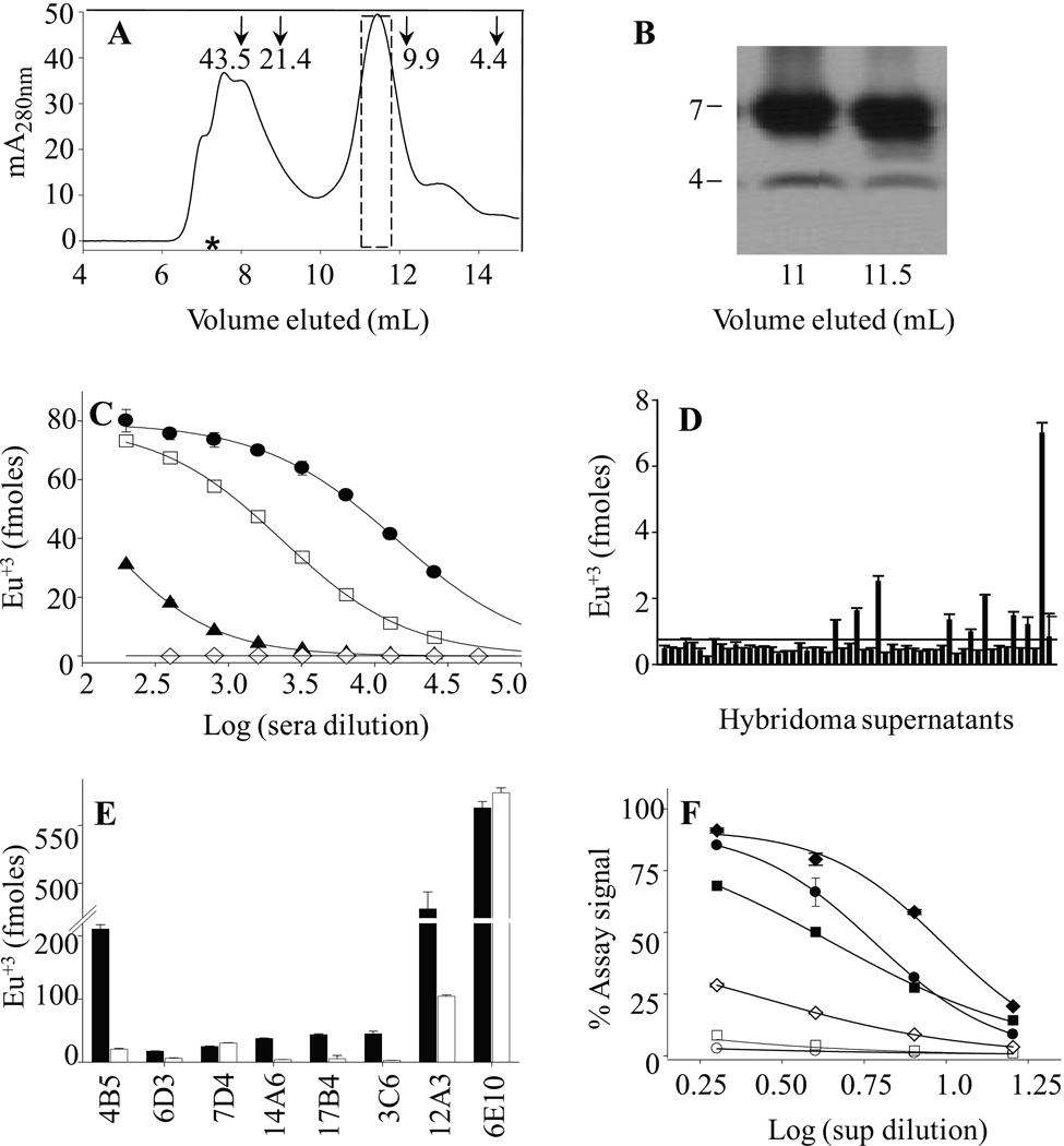
) samples on a Superdex™ 75 10/300-GL column eluted with 25 mM ammonium acetate, pH 8.5, confirmed their high purity and the absence of higher molecular weight Aβ assemblies. (F) Antibody binding curves for purified IgMκ's 3C6 (■, □) and 4B5 (▲,Δ), and control mAb 6E10 (♦, ◊) against plate-immobilized freshly isolated (Aβ1-40S26C)2 (closed symbols) and Aβ1-40S26C (open symbols). Antibody binding curves were also determined for 3C6 (G) and 4B5 (H) binding to: freshly-isolated (Aβ1-40S26C)2 (●); (Aβ1-40S26C)2 stored for ~1 month at −80 °C in 25 mM ammonium acetate, pH 8.5, (▼); Freshly-isolated Aβ1-40S26C monomers (□), and WT Aβ1-40 fibrils (◊). Antibody binding studies were carried out in PBSA containing 1% BSA and 0.05% Tween 20, pH 7.4.
Figure 3
Figure 3. Anti-Aβ dimer mAbs 3C6 and 4B5 binding to solution-phase Aβ conformers
Antibody binding to solution-phase Aβ conformers was established in a competition assay whereby mAbs 3C6 (A) and 4B5 (B) binding to plate-immobilized (Aβ1-40S26C)2 was determined in the presence of: freshly-isolated (Aβ1-40S26C)2 (●); (Aβ1-40S26C)2 stored for ~1 month at −80 °C in 25 mM ammonium acetate, pH 8.5, (▼); Freshly-isolated Aβ1-40S26C monomers (□), and WT Aβ1-40 fibrils (◊). Antibody binding was carried out in PBSA containing 1% BSA and 0.05% Tween 20, pH 7.4.
Figure 4
Figure 4. MAb 3C6 binds Aβ and ameliorates the block of LTP induced by AD brain extract
A TBS extract of AD brain was examined by immunoprecipitation (IP)/Western blotting using the polyclonal anti-Aβ antibody, AW8, for IP and a combination of anti-Aβ mAbs, 2G3 and 21F12, for Western blotting (A). The first two lanes of the Western blot show that the untreated TBS extract and buffer-exchanged extract contained highly similar amounts of Aβ monomer and SDS-stable Aβ dimers. The third lane shows that the first round of IP had effectively depleted the extract of all detectable Aβ. Molecular weight standards are indicated on the left, and Aβ monomers (M) and dimers (D) labeled with arrows on the right. Based on standard synthetic Aβ1-42 included on the blot, the test samples contained 50 ng/ml and 70 ng/ml Aβ dimer and monomer, respectively (B). Western blot of Aβ IP’d from AD brain TBS extract by the anti-Aβ polyclonal IgG, AW7; anti-IgM alone, and mAb 3C6 are shown in duplicate in lanes 1–6. To determine if 3C6 bound all the Aβ present in the AD TBS extract, the material not IP’d by 3C6 was used for IP with AW7 (lanes 7+8). Similarly, to determine the specificity of the bands detected by 3C6, this antibody was also used to IP a TBS extract from a non-demented control (lane 9). The blot shows that both 3C6 and AW7, but not the control IgM, IP’d Aβ assemblies that migrated on polyacrylamide gels as monomers and SDS-stable dimers. (C) Intracerebroventricular (i.c.v.) injection (*) of AD TBS brain extract inhibited high frequency stimulation (HFS, arrow) induced LTP (107 ± 5 %, n = 4, baseline, p>0.05 compared with pre-HFS baseline, and p<0.05 compared with vehicle injected controls (135 ± 5 %, n = 8)). (D) Co-injection of 5 µg of 3C6 prevented the inhibition of LTP by AD TBS extract (130 ± 5 %, n = 5, compared p<0.05 compared with AD TBS alone, n = 5), whereas co-injection of a control IgMκ, M1520, did not (105± 5 %, p<0.05, n = 5, compared with AD TBS extract + 3C6, p>0.05 compared with AD TBS extract alone). (E) LTP was not impaired when 10 µg of 3C6 (hash symbol) was i.c.v. injected 15 min before AD TBS brain extract (138 ± 5 % (n= 4) baseline, p<0.05 compared with 136 ± 6 % (n = 5) vehicle injected controls). Insets show representative EPSP traces at the times indicated. Calibration bars: vertical, 1 mV; horizontal, 10 ms.

Similar articles

Cited by

References

    1. Adekar SP, Klyubin I, Macy S, Rowan MJ, Solomon A, Dessain SK, O'Nuallain B. Inherent anti-amyloidogenic activity of human immunoglobulin gamma heavy chains. J Biol Chem. 2010;285:1066–1074. - PMC - PubMed
    1. Alzheimer A. Über einen eigenartigen schweren Erkrankungsproze! der Hirnrinde. Neurologisches Centralblatt. 1906;23:1129–1136.
    1. Arancio O, Chao MV. Neurotrophins, synaptic plasticity and dementia. Curr Opin Neurobiol. 2007;17:325–330. - PubMed
    1. Banks WA, Farr SA, Morley JE, Wolf KM, Geylis V, Steinitz M. Anti-amyloid beta protein antibody passage across the blood-brain barrier in the SAMP8 mouse model of Alzheimer's disease: an age-related selective uptake with reversal of learning impairment. Exp Neurol. 2007;206:248–256. - PMC - PubMed
    1. Barghorn S, Nimmrich V, Striebinger A, Krantz C, Keller P, Janson B, Bahr M, Schmidt M, Bitner RS, Harlan J, Barlow E, Ebert U, Hillen H. Globular amyloid beta-peptide oligomer - a homogenous and stable neuropathological protein in Alzheimer's disease. J Neurochem. 2005;95:834–847. - PubMed

Publication types

MeSH terms