

Reactivation of latent tuberculosis in rhesus macaques by coinfection with simian immunodeficiency virus
- PMID: 21781131
- PMCID: PMC3227019
- DOI: 10.1111/j.1600-0684.2011.00485.x
Reactivation of latent tuberculosis in rhesus macaques by coinfection with simian immunodeficiency virus
Abstract
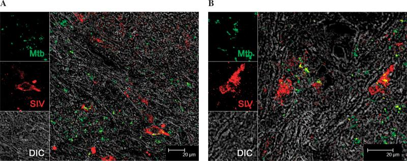
Background: Tuberculosis (TB) and AIDS together present a devastating public health challenge. Over 3 million deaths every year are attributed to these twin epidemics. Annually, ∼11 million people are coinfected with HIV and Mycobacterium tuberculosis (Mtb). AIDS is thought to alter the spontaneous rate of latent TB reactivation.
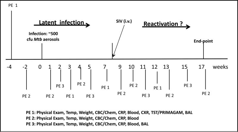
Methodology: Macaques are excellent models of both TB and AIDS. Therefore, it is conceivable that they can also be used to model coinfection. Using clinical, pathological, and microbiological data, we addressed whether latent TB infection in rhesus macaques can be reactivated by infection with simian immunodeficiency virus (SIV).
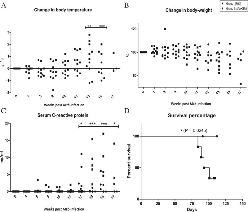
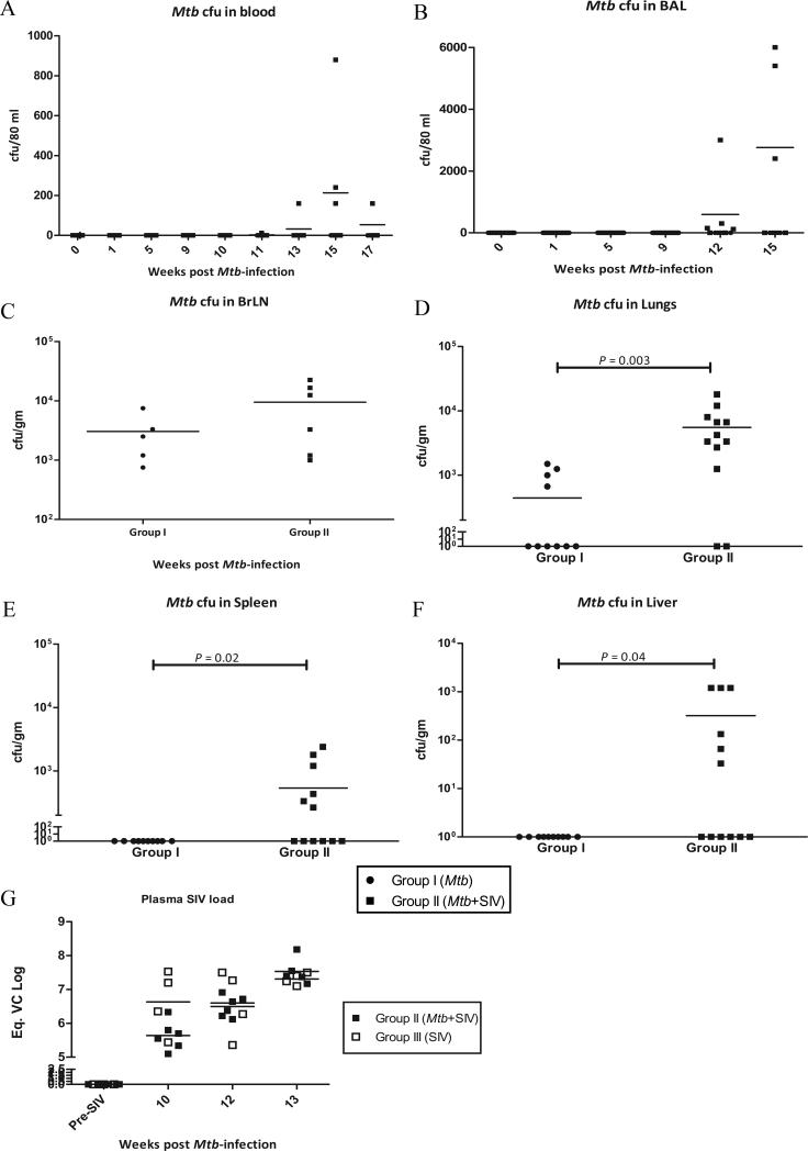
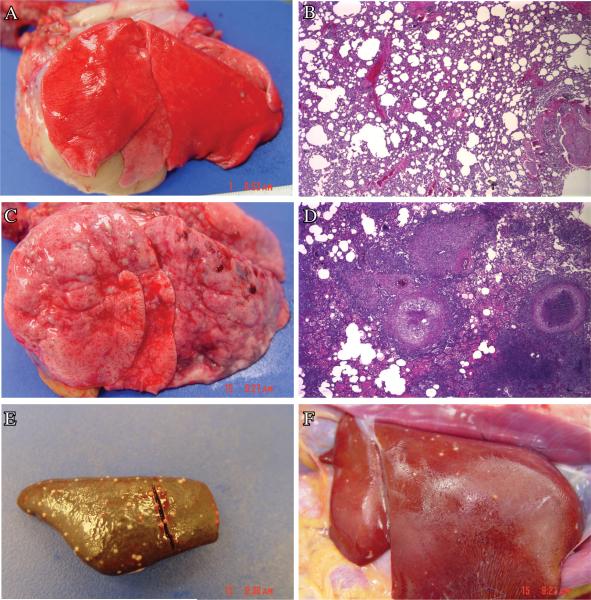
Results: A low-dose aerosol infection of rhesus macaques with Mtb caused latent, asymptomatic TB infection. Infection of macaques exhibiting latent TB with a rhesus-specific strain of SIV significantly reactivated TB.
Conclusions: Rhesus macaques are excellent model of TB/AIDS coinfection and can be used to study the phenomena of TB latency and reactivation.
© 2011 John Wiley & Sons A/S.
Figures
Similar articles
-
High Turnover of Tissue Macrophages Contributes to Tuberculosis Reactivation in Simian Immunodeficiency Virus-Infected Rhesus Macaques.J Infect Dis. 2018 May 25;217(12):1865-1874. doi: 10.1093/infdis/jix625. J Infect Dis. 2018. PMID: 29432596 Free PMC article.
-
Simian immunodeficiency virus-induced changes in T cell cytokine responses in cynomolgus macaques with latent Mycobacterium tuberculosis infection are associated with timing of reactivation.J Immunol. 2011 Mar 15;186(6):3527-37. doi: 10.4049/jimmunol.1003773. Epub 2011 Feb 11. J Immunol. 2011. PMID: 21317393 Free PMC article.
-
Antiretroviral therapy does not reduce tuberculosis reactivation in a tuberculosis-HIV coinfection model.J Clin Invest. 2020 Oct 1;130(10):5171-5179. doi: 10.1172/JCI136502. J Clin Invest. 2020. PMID: 32544085 Free PMC article.
-
Chronic Immune Activation in TB/HIV Co-infection.Trends Microbiol. 2020 Aug;28(8):619-632. doi: 10.1016/j.tim.2020.03.015. Epub 2020 Apr 22. Trends Microbiol. 2020. PMID: 32417227 Free PMC article. Review.
-
Monkeying around with MAIT Cells: Studying the Role of MAIT Cells in SIV and Mtb Co-Infection.Viruses. 2021 May 8;13(5):863. doi: 10.3390/v13050863. Viruses. 2021. PMID: 34066765 Free PMC article. Review.
Cited by
-
Concurrent TB and HIV therapies effectively control clinical reactivation of TB during co-infection but fail to eliminate chronic immune activation.Res Sq [Preprint]. 2024 Aug 26:rs.3.rs-4908400. doi: 10.21203/rs.3.rs-4908400/v1. Res Sq. 2024. PMID: 39257997 Free PMC article. Preprint.
-
Immunology studies in non-human primate models of tuberculosis.Immunol Rev. 2015 Mar;264(1):60-73. doi: 10.1111/imr.12258. Immunol Rev. 2015. PMID: 25703552 Free PMC article. Review.
-
Immune correlates of tuberculosis disease and risk translate across species.Sci Transl Med. 2020 Jan 29;12(528):eaay0233. doi: 10.1126/scitranslmed.aay0233. Sci Transl Med. 2020. PMID: 31996462 Free PMC article.
-
Mucosal vaccination with attenuated Mycobacterium tuberculosis induces strong central memory responses and protects against tuberculosis.Nat Commun. 2015 Oct 13;6:8533. doi: 10.1038/ncomms9533. Nat Commun. 2015. PMID: 26460802 Free PMC article.
-
Systems biology approaches to investigate the role of granulomas in TB-HIV coinfection.Front Immunol. 2022 Oct 31;13:1014515. doi: 10.3389/fimmu.2022.1014515. eCollection 2022. Front Immunol. 2022. PMID: 36405707 Free PMC article. Review.
References
-
- Dye C, Scheele C, Dolin P, Pathania V, Raviglione MC. Global burden of tuberculosis: estimated incidence, prevalence, and mortality by country. JAMA. 1999;282:677–686. - PubMed
-
- Centers for Disease Control and Prevention Prevention and treatment of tuberculosis among patients infected with human immunodeficiency virus: principles of therapy and revised recommendations. Morb Mortal Wkly Rep. 1998;47(RR-20):1–58. - PubMed
-
- Joint United Nations Programme on AIDS (UNAIDS) WHO . AIDS Epidemic Update Rep. 02.46E 2002. UNAIDS/WHO; Geneva:
-
- Steinbrook R. One Step Forward, Two Steps Back — Will There Ever Be an AIDS Vaccine? New Engl J Med. 2007;357:2653–2655. - PubMed
-
- Corbett EL, Watt CJ, Walker N, Maher D, Williams BG, Raviglione MC, Dye C. The growing burden of tuberculosis: global trends and interactions with the HIV epidemic. Arch Intern Med. 2003;163:1009–21. - PubMed
Publication types
MeSH terms
Substances
Grants and funding
- HL10679/HL/NHLBI NIH HHS/United States
- R01 HL106790/HL/NHLBI NIH HHS/United States
- P51 RR000164/RR/NCRR NIH HHS/United States
- RR020159/RR/NCRR NIH HHS/United States
- RR026006/RR/NCRR NIH HHS/United States
- AI089323/AI/NIAID NIH HHS/United States
- R01 AI089323/AI/NIAID NIH HHS/United States
- RR000164/RR/NCRR NIH HHS/United States
- P20 RR020159/RR/NCRR NIH HHS/United States
- AI084793/AI/NIAID NIH HHS/United States
- R21 RR026006/RR/NCRR NIH HHS/United States
- R21 AI091457/AI/NIAID NIH HHS/United States
- AI091457/AI/NIAID NIH HHS/United States
- R01 AI084793/AI/NIAID NIH HHS/United States
LinkOut - more resources
Full Text Sources
