

Increased plasma chymase concentration and mast cell chymase expression in venous neointimal lesions of patients with CKD and ESRD
- PMID: 21781173
- PMCID: PMC3212616
- DOI: 10.1111/j.1525-139X.2011.00921.x
Increased plasma chymase concentration and mast cell chymase expression in venous neointimal lesions of patients with CKD and ESRD
Abstract
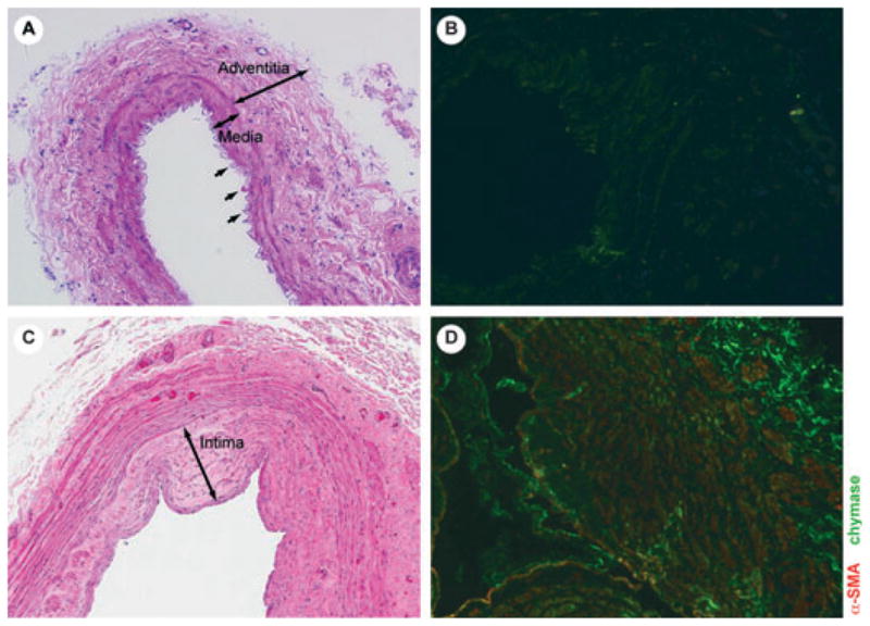
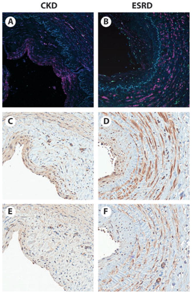
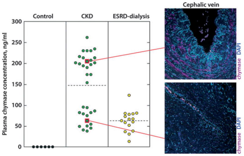
The underlying inflammatory component of chronic kidney disease may predispose blood vessels to intimal hyperplasia (IH), which is the primary cause of dialysis access failure. We hypothesize that vascular pathology and markers of IH formation are antecedent to arteriovenous (AV) fistula creation. Blood, cephalic, and basilic vein segments were collected from predialysis chronic kidney disease (CKD) patients with no previous AV access and patients with end-stage renal disease (ESRD). Immunohistochemistry was performed with antibodies against mast cell chymase, transforming growth factor-beta (TGF-β) and interleukin-6 (IL-6), which cause IH. Plasma chymase was measured by ELISA. IH was present in 91% of CKD and 75% of ESRD vein segments. Chymase was abundant in vessels with IH, with the greatest expression in intima and medial layers, and virtually absent in the controls. Chymase colocalized with TGF-β1 and IL-6. Plasma chymase concentration was elevated up to 33-fold in patients with CKD versus controls and was associated with increased chymase in vessels with IH. We show that chymase expression in vessels with IH corresponds with plasma chymase concentrations. As chymase inhibition attenuates IH in animal models, and we find chymase is highly expressed in IH lesions of patients with CKD and ESRD, we speculate that chymase inhibition could have therapeutic value in humans.
© 2011 Wiley Periodicals, Inc.
Figures
References
-
- Roy-Chaudhury P, Arend L, Zhang J, Krishnamoorthy M, Wang Y, Banerjee R, Samaha A, Munda R. Neointimal hyperplasia in early arteriovenous fistula failure. Am J Kidney Dis. 2007;50:782–790. - PubMed
-
- Roy-Chaudhury P, Kelly BS, Miller MA, Reaves A, Armstrong J, Nanayakkara N, Heffelfinger SC. Venous neointimal hyperplasia in polytetrafluoroethylene dialysis grafts. Kidney Int. 2001;59:2325–2334. - PubMed
-
- Beaulieu MC, Gabana C, Rose C, MacDonald PS, Clement J, Kiaii M. Stenosis at the area of transposition—an under-recognized complication of transposed brachiobasilic fistulas. J Vasc Access. 2007;8:268–274. - PubMed
-
- Quinn SF, Schuman ES, Hall L, Gross GF, Uchida BT, Standage BA, Rosch J, Ivancev K. Venous stenoses in patients who undergo hemodialysis: treatment with self-expandable endovascular stents. Radiology. 1992;183:499–504. - PubMed
-
- Vorwerk D, Guenther RW, Mann H, Bohndorf K, Keulers P, Alzen G, Sohn M, Kistler D. Venous stenosisocclusion in hemodialysis shunts: follow-up results of stent placement in 65 patients. Radiology. 1995;195:140–146. - PubMed
Publication types
MeSH terms
Substances
Grants and funding
LinkOut - more resources
Full Text Sources
Other Literature Sources
Medical
Research Materials
