

Review
doi: 10.1111/j.1600-0757.2011.00386.x.
Squamous cell carcinoma and precursor lesions: diagnosis and screening in a technical era
- PMID: 21781180
- PMCID: PMC3145964
- DOI: 10.1111/j.1600-0757.2011.00386.x
Item in Clipboard
Review
Squamous cell carcinoma and precursor lesions: diagnosis and screening in a technical era
Periodontol 2000.
2011 Oct.
No abstract available
Figures
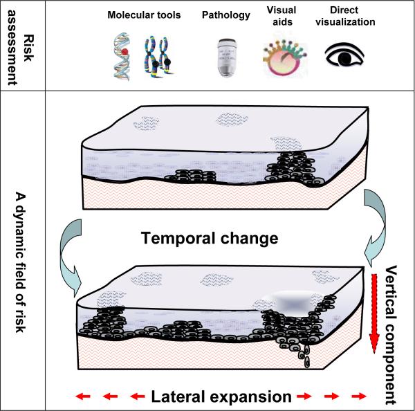
Assessment of a dynamic field at risk
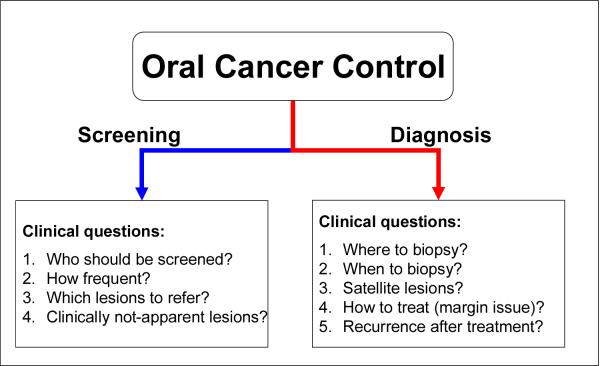
Clinical questions in oral cancer control in screening and diagnosis
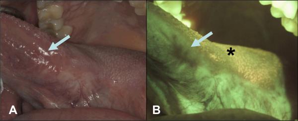
A 42-year-old male nonsmoker was examined 4 years after surgery and radiation therapy for Stage III squamous cell carcinoma involving the left lateral tongue and left cervical lymph nodes. A. White light image of a well-healed scar at left lateral tongue with no clinically visible lesion (arrow); B. The same area (arrow) under FV showing a dark brown FVL. The comparative biopsy from the FVL area 4-and-half years after initial treatment showed severe epithelial dysplasia. The area posterior to the FVL showed FVR representing a well-healed scar (star).
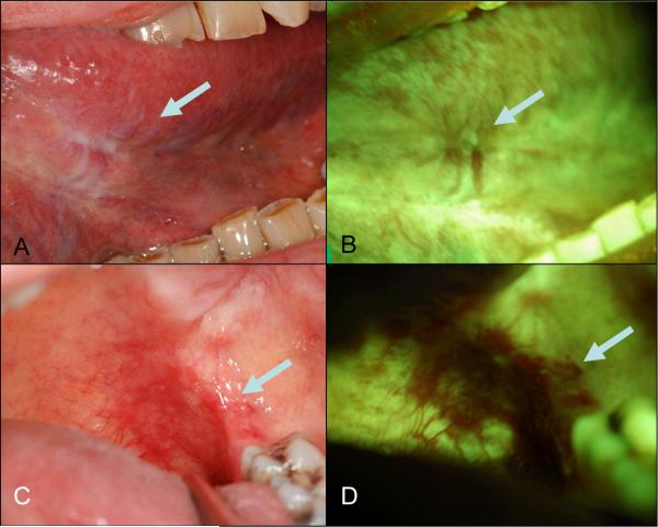
A 68-year-old female smoker presented with a carcinoma in situ on the left anterior ventral tongue after completion of an excisional biopsy with margin positive for mild epithelial dysplasia. At 3 months post surgery, she was referred for the assessment of the surgical site. A. White light image showing a scar without clinically visible lesion on left anterior ventral tongue (arrow); B. FV image showing some FVL area (arrow). C. White light image showing an ill-defined mildly erythematous area at left posterior soft palate and FV image showing a well-defined dark brown FVL area (D, arrow) 8 cm distant to the initial cancer site. The biopsy showed carcinoma in situ.
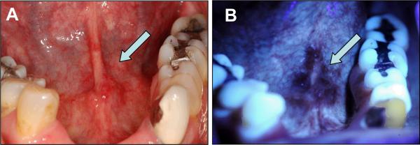
A 55-year-old female former smoker presented with a pathology-proven severe epithelial dysplasia at the anterior of floor of mouth extending to the lingual frenum. A. White light image showing an ill-defined, slightly erythematous lesion (arrow) at anterior of mouth; B. FV image showing a well-demarcated area of FVL area (arrow).
Similar articles
-
Oral cancer: the role of the dentist in prevention and early detection.Dent Today. 2001 May;20(5):92-7. Dent Today. 2001. PMID: 11443823 No abstract available.
-
New initiatives in the diagnosis and prevention of oral cancer.Northwest Dent. 2004 Jul-Aug;83(4):21-2, 24-6, 28 passim. Northwest Dent. 2004. PMID: 15462557 No abstract available.
-
Diagnostic devices for detecting oral cancer.J Dent Hyg. 2009 Fall;83(4):177-8. Epub 2009 Nov 2. J Dent Hyg. 2009. PMID: 19909635 No abstract available.
-
Squamous cell carcinoma and precursor lesions: prevention.Periodontol 2000. 2011 Oct;57(1):38-50. doi: 10.1111/j.1600-0757.2011.00395.x. Periodontol 2000. 2011. PMID: 21781178 Review. No abstract available.
-
Screening for oral potentially malignant epithelial lesions and squamous cell carcinoma: a discussion of benefit and risk.J Can Dent Assoc. 2014;80:e47. J Can Dent Assoc. 2014. PMID: 25055237 Review. No abstract available.
Cited by
-
Influence of fluorescence on screening decisions for oral mucosal lesions in community dental practices.J Oral Pathol Med. 2014 Jan;43(1):7-13. doi: 10.1111/jop.12090. Epub 2013 Jun 10. J Oral Pathol Med. 2014. PMID: 23750637 Free PMC article.
-
FDI policy statement on oral cancer: Adopted by the FDI General Assembly: 24 September 2015, Bangkok, Thailand.Int Dent J. 2016 Feb;66(1):13-4. doi: 10.1111/idj.12234. Int Dent J. 2016. PMID: 26803944 Free PMC article. No abstract available.
-
Targeting of chemoprevention to high-risk potentially malignant oral lesions: challenges and opportunities.Oral Oncol. 2014 Dec;50(12):1123-30. doi: 10.1016/j.oraloncology.2014.08.012. Epub 2014 Sep 16. Oral Oncol. 2014. PMID: 25240917 Free PMC article.
-
In vivo wide-field reflectance/fluorescence imaging and polarization-sensitive optical coherence tomography of human oral cavity with a forward-viewing probe.Biomed Opt Express. 2015 Jan 14;6(2):524-35. doi: 10.1364/BOE.6.000524. eCollection 2015 Feb 1. Biomed Opt Express. 2015. PMID: 25780742 Free PMC article.
-
Accuracy of in vivo multimodal optical imaging for detection of oral neoplasia.Cancer Prev Res (Phila). 2012 Jun;5(6):801-9. doi: 10.1158/1940-6207.CAPR-11-0555. Epub 2012 May 2. Cancer Prev Res (Phila). 2012. PMID: 22551901 Free PMC article.
References
-
- Banda-Gamboa H, Ricketts I, Cairns A, Hussein K, Tucker JH, Husain N. Automation in cervical cytology: an overview. Anal Cell Pathol. 1992;4:25–48. - PubMed
-
- Barrellier P, Babin E, Louis MY, Meunier-Guttin A. The use of toluidine blue in the diagnosis of neoplastic lesions of the oral cavity. Rev Stomatol Chir Maxillofac. 1993;94:51–54. - PubMed
-
- Bocking A, Nguyen VQ. Diagnostic and prognostic use of DNA image cytometry in cervical squamous intraepithelial lesions and invasive carcinoma. Cancer. 2004;102:41–54. - PubMed
-
- Brennan JA, Mao L, Hruban RH, Boyle JO, Eby YJ, Koch WM, Goodman SN, Sidransky D. Molecular assessment of histopathological staging in squamous-cell carcinoma of the head and neck. N Engl J Med. 1995;332:429–435. - PubMed
-
- Califano J, van der Riet P, Westra W, Nawroz H, Clayman G, Piantadosi S, Corio R, Lee D, Greenberg B, Koch W, Sidransky D. Genetic progression model for head and neck cancer: implications for field cancerization. Cancer Res. 1996;56:2488–2492. - PubMed
Publication types
MeSH terms
Substances
Grants and funding
LinkOut - more resources
Full Text Sources
Medical
