Review
. 2011 Jul;9 Suppl 1(0 1):130-43.
doi: 10.1111/j.1538-7836.2011.04320.x.

Biology and physics of von Willebrand factor concatamers

Affiliations
Review

Biology and physics of von Willebrand factor concatamers

T A Springer. J Thromb Haemost. 2011 Jul.

Abstract

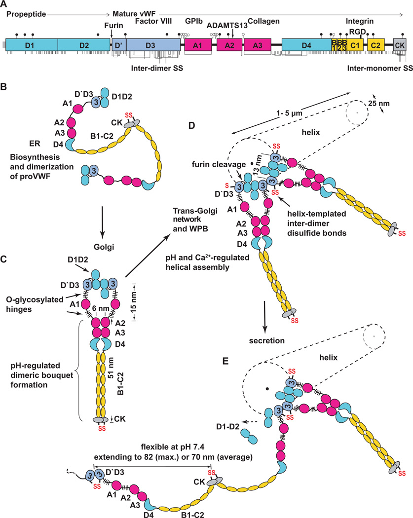
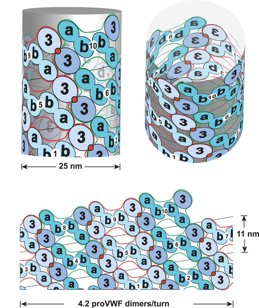
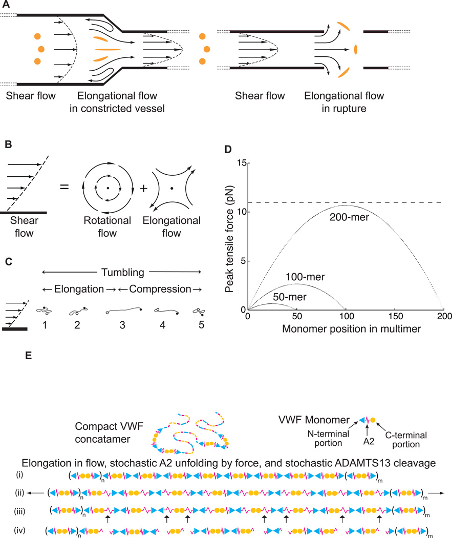
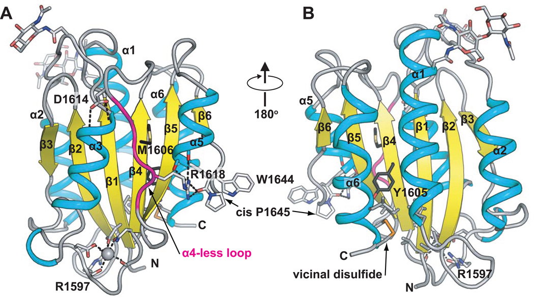
Structural specialisations enable von Willebrand factor (VWF) to assemble during biosynthesis into helical tubules in Weibel-Palade bodies (WPB). Specialisations include a pH-regulated dimeric bouquet formed by the C-terminal half of VWF and helical assembly guided by the N-terminal half that templates inter-dimer disulphide bridges. Orderly assembly and storage of ultra-long concatamers in helical tubules, without crosslinking of neighboring tubules, enables unfurling during secretion without entanglement. Length regulation occurs post-secretion, by hydrodynamic force-regulated unfolding of the VWF A2 domain, and its cleavage by the plasma protease ADAMTS13 (a disintegrin and metalloprotease with a thrombospondin type 1 motif, member 13). VWF is longest at its site of secretion, where its haemostatic function is most important. Moreover, elongational hydrodynamic forces on VWF are strongest just where needed, when bound to the vessel wall, or in elongational flow in the circulation at sites of vessel rupture or vasoconstriction in haemostasis. Elongational forces regulate haemostasis by activating binding of the A1 domain to platelet GPIbα, and over longer time periods, regulate VWF length by unfolding of the A2 domain for cleavage by ADAMTS13. Recent structures of A2 and single molecule measurements of A2 unfolding and cleavage by ADAMTS13 illuminate the mechanisms of VWF length regulation. Single molecule studies on the A1-GPIb receptor-ligand bond demonstrate a specialised flex-bond that enhances resistance to the strong hydrodynamic forces experienced at sites of haemorrhage.


Figures

Figure 1
Figure 1
Biosynthesis, helical assembly, and secretion of VWF concatamers. A. Primary structure and domain organisation of VWF [66]. Cysteines are vertical lines and are connected for chemically determined disulphide bonds [67, 68]. N and O-linked glycans are closed and open lollipops, respectively [9]. B–E. Scheme for biosynthesis, helical assembly, and secretion. EM evidence for C-terminal pH-regulated dimeric bouquet and domain shapes is from Y.-F. Zhou, E., Eng, N. Nishida, C. Lu, T. Walz, and T.A. Springer, unpublished. EM evidence for N-terminal D1–D2 homodimerisation and the helical assembly is from [8].
Figure 2
Figure 2
Helical assembly of the N-terminal domains of VWF in the tubules of WPB. Modified from [8]. Helical assembly is shown as progressing from bottom to top. Each successive proVWF dimer is numbered and shown alternately as lighter domains outlined in red or darker outlined in green. The density for the D’D3 domains (3) is assigned based on larger size than the D1 and D2 domains (a and b). Inter-dimer disulphide crosslinks form at the 2-fold symmetry axis between D’D3 domains (red circles). The D1 and D2 domains are denoted a and b since it is not known which is in which position in the helical assembly. Noncovalent, homomeric interactions within each dimer are mediated by either the D1 or D2 domain, in the b position [8].
Figure 3
Figure 3
Shear and elongational flow, VWF concatamer conformation, and mechanoenzymatic cleavage. Modified from [20]. A. Shear flow, and transition to elongational flow in vessels at sites of constricted or ruptured vessels. Round orange spheres show the effect of enlongational flow on the shape of a polymeric protein in the flow field (adapted from [21]). Elongation of VWF concatamers would occur in the indicated directions. B. Shear flow, which may be represented as elongational flow superimposed on rotational flow [22]. Arrows show stream lines and dots regions of no flow. C. Cartoon of VWF elongating, compressing, and tumbling in shear flow. D. Peak force as function of monomer position in a VWF multimer chain of 200, 100 or 50 monomers at 100 dyn/cm2. Dashed line shows the most likely unfolding force for the A2 domain at a loading rate of 25 pN/s. E. Schematic of VWF, with N-terminal end as triangle, A2 as spring, and C-terminal end as circle. Elongation results in stochastic unfolding of some A2 domains (ii), some of which are cleaved by ADAMTS13 (iii). The resulting fragments are shown (iv).
Figure 4
Figure 4
The VWF A2 domain. Ribbon diagrams show α-helices (cyan), β-strands (yellow) and loops (grey). The α4-less loop is in magenta. Key sidechains are shown as sticks, and Ca2+ as a sphere. Carbons of the ADAMTS13 cleavage site residues Tyr1605 and Met1606 are black. The vicinal disulphide is in gold. Two Asn-linked carbohydrates are in stick near the top of the domain. N and C-termini are marked. Hydrogen and metal coordination bonds are dashed. The chimeric model was made by superimposing two A2 structures [30, 31], and grafting the Ca2+-bound α3-β loop [31] onto the mammalian wild type structure [30].
Figure 5
Figure 5
Single molecule demonstration of A2 domain unfolding, refolding, and mechanoenzymatic cleavage. Modified from [47]. A. Schematic diagram of the DNA handle-A2 domain construct (upper) and laser trap (lower). The A2 domain cartoon is in rainbow, blue to red from N-terminus to C-terminus, respectively. The A2 domain is coupled through Cys-Gly-Gly and Gly-Gly-Cys linkages and disulphides to double-stranded DNA handles, that have biotin and digoxygenin tags at the opposite ends. The tether is held by beads functionalised with streptavidin and digoxigenin Fab. Force is applied by micropipette movement (right), and measured by bead displacement in the laser trap (left). B. Three representative cycles of force increase, decrease, and clamping at a constant low level. C. Traces of force versus tether extension in the force increase phases of cycles ii and iii in B. An abrupt unfolding event is seen in ii and not iii. It is inferred that A2 was unfolded at the beginning of iii. D. Representative traces showing ADAMTS13 cleavage (1) and no cleavage (2). In each trace unfolding of A2 is seen, and A2 is returned to a clamped force of 5 pN to assess kinetics of cleavage. Cleavage of the tether returns force to 0. A new tether must be formed for each test of cleavage; cleavage is measured one A2 molecule at a time.
Figure 6
Figure 6
Repeated measurement of GPIbα and VWF A1 domain binding and unbinding in a single molecule. Modified from [47]. A. Schematic. The A1 domain and GPIbα complex crystal structure are shown as ribbon diagrams, with native disulphides in gold. The ReaLiSM fusion construct contains from N to C the A1 domain, a 43 or 26-residue polypeptide linker, and GPIbα, and is expressed as a secreted protein in mammalian cells. Cysteines are included at the N and C-termini for disulphide linkage to DNA handles, which are coupled to beads as in Fig. 5. The mini-laser tweezers differs from that in Fig. 5. Force is applied by movement of the laser trap, and measured as a change in light momentum. B. One representative cycle. Unbinding and rebinding are measured as abrupt changes in tether extension during pulling (black) and relaxation (red). C. Kinetic measurements. Data from each bin in E–H is a different symbol, and each symbol shape/fill is at a different pulling rate. Red diamonds are from hopping kinetics in force-clamp mode [47]. Open circle at 0 pN is bulk phase data from 125I-A1 domain dissociation from platelets [55]. D. Schematic model of a flex-bond. A1 (cyan) and GPIbα (magenta) are subjected to tensile force at the N and C termini of the ReaLiSM construct (arrows). k1off and k2off are dissociation rates from two different states. k21 and k12 are rates of equilibration between the two bound states. Measured dissociation kinetics reflect all four kinetic rates. E–H. Unbinding force distributions at four different indicated pulling rates. Dissociation at a higher force occurs at faster pulling rates. As predicted by theory and global fit of all data (red curves), the average force at which each pathway dissociates increases slightly with loading rate. I and J. Unbinding force distributions at the faster pulling rate in presence of ristocetin and botrocetin.
Figure 7
Figure 7
Crystal structure of the complex of GPIbα LRR domain and VWF A1, with mutations marked [53, 54]. Disulphides are shown with gold spheres. VWD type 2B mutations in A1 and platelet-type VWD mutations in GPIbα are shown with Cα atom spheres and ribbon in red and green, respectively. All mutations are gain-of-function, i.e. they increase adhesiveness. The A1 mutations map to regions that are buried and in the vicinity of the long-range disulphide bond, and are mostly not in contact with GPIbα. The GPIbα mutations are in its β-thumb, which changes conformation when bound to A1. LRR 2–4 of GPIbα are in grey. They are not in contact with A1 in the crystal structure, and are not important in binding to A1 in stasis, but become increasingly required for binding to A1 as shear is increased, as shown by exchange for canine sequence [64].

References

    1. Sadler JE. New concepts in von Willebrand disease. Annu Rev Med. 2005;56:173–191. - PubMed
    1. Wagner DD. Cell biology of von Willebrand factor. Annu Rev Cell Biol. 1990;6:217–246. - PubMed
    1. Sadler JE. Biochemistry and genetics of von Willebrand factor. Annu Rev Biochem. 1998;67:395–424. - PubMed
    1. Ruggeri ZM, Mendolicchio GL. Adhesion mechanisms in platelet function. Circ Res. 2007;100:1673–1685. - PubMed
    1. Zenner HL, Collinson LM, Michaux G, Cutler DF. High-pressure freezing provides insights into Weibel-Palade body biogenesis. J Cell Sci. 2007;120:2117–2125. - PubMed

Publication types

Substances