. 2011 Jul 23;10(1):4.
doi: 10.1186/1476-5926-10-4.

Characterization of chronic HCV infection-induced apoptosis

Affiliations

Characterization of chronic HCV infection-induced apoptosis

Abdel-Rahman N Zekri et al. Comp Hepatol. .

Abstract

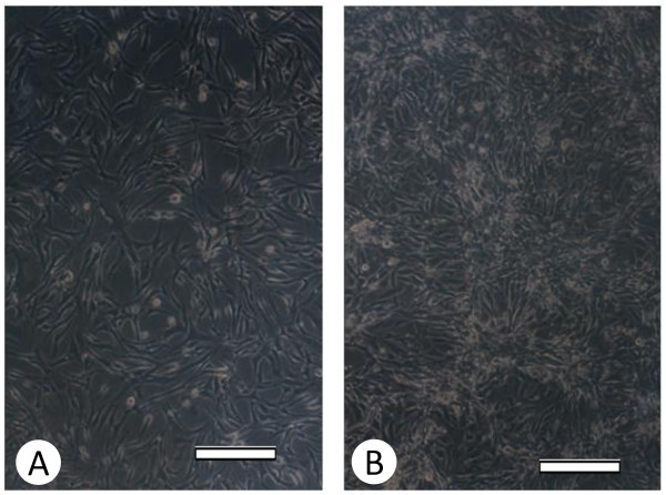
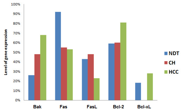
Background: To understand the complex and largely not well-understood apoptotic pathway and immune system evasion mechanisms in hepatitis C virus (HCV)-associated hepatocellular carcinoma (HCC) and HCV associated chronic hepatitis (CH), we studied the expression patterns of a number of pro-apoptotic and anti-apoptotic genes (Fas, FasL, Bcl-2, Bcl-xL and Bak) in HepG2 cell line harboring HCV- genotype-4 replication. For confirmation, we also assessed the expression levels of the same group of genes in clinical samples obtained from 35 HCC and 34 CH patients.

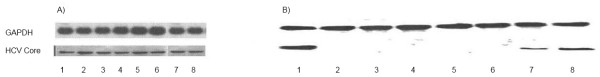
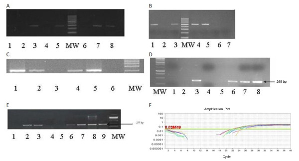
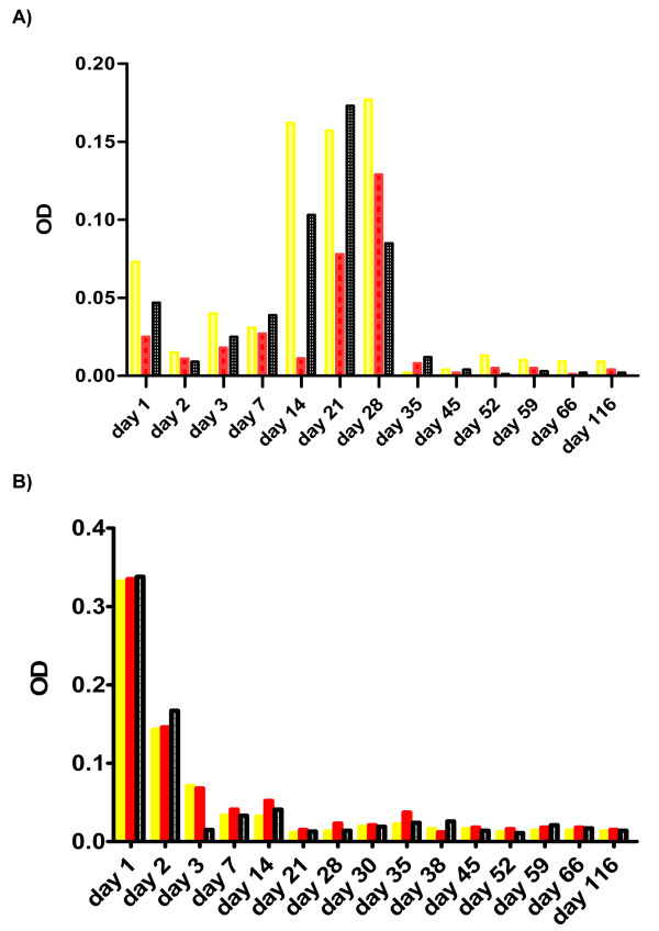
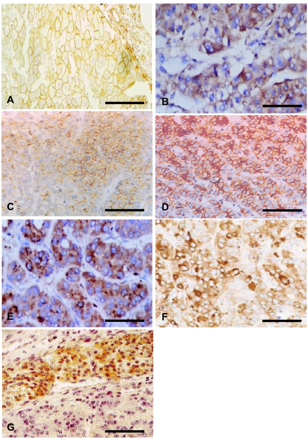
Methods: Viral replication was assessed in the tissue culture medium by RT-PCR, quantitative Real-Time PCR (qRT-PCR); detection of HCV core protein by western blot and inhibition of HCV replication with siRNA. The expression level of Fas, FasL, Bcl-2, Bcl-xL and Bak was assessed by immunohistochemistry and RT-PCR whereas caspases 3, 8 and 9 were assessed by colorimetric assay kits up to 135 days post infection.

Results: There was a consistent increase in apoptotic activity for the first 4 weeks post-CV infection followed by a consistent decrease up to the end of the experiment. The concordance between the changes in the expression levels of Fas, FasL, Bcl-2, Bcl-xL and Bak in vitro and in situ was statistically significant (p < 0.05). Fas was highly expressed at early stages of infection in cell lines and in normal control liver tissues followed by a dramatic reduction post-HCV infection and an increase in the expression level of FasL post HCV infection. The effect of HCV infection on other apoptotic proteins started very early post-infection, suggesting that hepatitis C modulating apoptosis by modulating intracellular pro-apoptotic signals.

Conclusions: Chronic HCV infection differently modulates the apoptotic machinery during the course of infection, where the virus induces apoptosis early in the course of infection, and as the disease progresses apoptosis is modulated. This study could open a new opportunity for understanding the various signaling of apoptosis and in the developing a targeted therapy to inhibit viral persistence and HCC development.


Figures

Figure 1
Figure 1
(A): Non-infected HePG2 cells. (B): Infected HePG2 cells. Scale bar = 100 μm.
Figure 2
Figure 2
Expression levels of the viral core and GAPDH. (A) The expression level of the viral core and GAPDH in HepG2 cells infected by HCV genotype-4 from day 1 to day 8. (B) The expression level of the viral core in HepG-2 cells infected by HCV genotype-4 from day 1 to day 8. Upper row show HCV-core expression in un-transfected cells. Lower row showed the HCV- core expression in siRNA-Z5 transfected cells.
Figure 3
Figure 3
Data on gene amplification. Ethidium bromide-stained 2% agarose gel (A) for Bcl2 gene amplification. Lanes 1 and 2 showed negative RT-PCR control; lane 3 showed positive amplification of CH case; lane 4 showed negative amplification of CH case; lane 5 showed positive amplification of HCC case; lane 6 showed negative amplification of HCC case; lane 7 showed positive amplification of HepG2 without HCV infection; lane 8 showed positive amplification of HepG2 with HCV infection. (B) For Bcl-Xl gene amplification. Lane 1 showed HepG2-positive amplification with HCV infection at day 28; lane 2 HepG2-negative amplification without HCV infection; lane 3 and 4 showed positive amplification of CH case; lane 5 showed positive amplification of HCC case; lane 6 & 7 showed negative RT-PCR control. (C) For Bak gene amplification. lane 1 HepG2-positive amplification with HCV infection at days 59; lane 2 HepG2-negative amplification without HCV infection lane 3 showed HepG2-negative amplification with HCV infection at days 35; lane 4 showed positive amplification of CH case; lane 5 showed positive amplification of HCC case of CH; lane 6 negative RT-PCR control. (D) for Fas gene amplification, first lane: MW, lanes 1 and 2: negative RT-PCR control, lane 3 showed HepG2-positive amplification without HCV infection, lane 4 HepG2- showed negative amplification with HCV infection at day 21, lane 5 showed negative case of HCC, lanes 6 and 7 showed positive amplification of CH and lane 8 showed positive amplification of HCC case. (E) for FasL gene amplification, lane 1: negative RT-PCR control; lanes 2 and 3 showed HepG2-positive amplification with HCV infection at days 28 and 35 respectively; lane 4 showed HepG2-negative amplification without HCV infection; lane 5 showed negative case of CH; lanes 6 and 7 showed positive amplification of CH, lanes 8 and 9 showed positive amplification of HCC case. (F) Amplification plot of RT-PCR for housekeeping gene using Taqman probe.
Figure 4
Figure 4
Changes in caspases expression levels in vitro.
Figure 5
Figure 5
The expression level of the apoptotic genes in the different studied groups. NB: CH = Chronic hepatitis, HCC = Hepatocelullar carcinoma, NAT = Normal distant to tumor.
Figure 6
Figure 6
Cases of chronic hepatitis (CH) and hepatocellular carcinoma (HCC). Data from cases of CH showing (A) high membranous expression of FasL, (B) moderate cytoplasmic expression of FAS and (C) moderate cytoplasmic expression of Bcl-2. Cases of HCC showing (D) High membranous expression of FasL, (E) Marked expression of FAS, (F) high expression of Bcl-2, and (G) Marked expression of Bcl2 in tumor tissues with loss of expression in adjacent non neoplastic region. Scale bar = 100 μm (A, C, D, G) and 200 μm (B, E, F).

Similar articles

Cited by

References

    1. Shepard CW, Finelli L, Alter MJ. Global epidemiology of hepatitis C virus infection. Lancet Infect Dis. 2005;5(9):558–567. doi: 10.1016/S1473-3099(05)70216-4. - DOI - PubMed
    1. Eassa S, Eissa M, Sharaf SM, Ibrahim MH, Hassanein OM. Prevalence of hepatitis C virus infection and evaluation of a health education program in el-ghar village in zagazig, egypt. J Egypt Public Health Assoc. 2007;82(5-6):379–404. - PubMed
    1. AbdulQawi K, Youssef A, Metwally MA, Ragih I, AbdulHamid M, Shaheen A. Prospective study of prevalence and risk factors for hepatitis C in pregnant Egyptian women and its transmission to their infants. Croat Med J. 2010;51(3):219–228. doi: 10.3325/cmj.2010.51.219. - DOI - PMC - PubMed
    1. El-Karaksy HM, Anwar G, Esmat G, Mansour S, Sabry M, Helmy H, El-Hennawy A, Fouad H. Prevalence of hepatic abnormalities in a cohort of Egyptian children with type 1 diabetes mellitus. Pediatr Diabetes. 2009;1(7):462–70. - PubMed
    1. Fischer R, Baumert T, Blum HE. Hepatitis C virus infection and apoptosis. World J Gastroenterol. 2007;13(36):4865–4872. - PMC - PubMed

LinkOut - more resources