. 2011 Nov;127(3-5):358-66.
doi: 10.1016/j.jsbmb.2011.07.002. Epub 2011 Jul 14.

Identification of a GPER/GPR30 antagonist with improved estrogen receptor counterselectivity

Affiliations

Identification of a GPER/GPR30 antagonist with improved estrogen receptor counterselectivity

Megan K Dennis et al. J Steroid Biochem Mol Biol. 2011 Nov.

Abstract

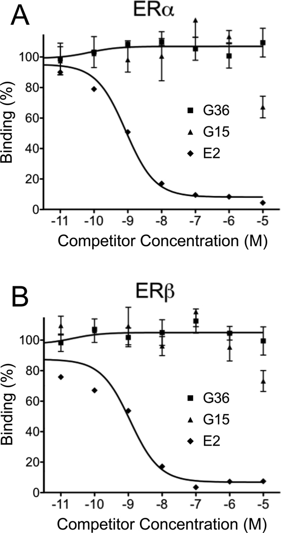
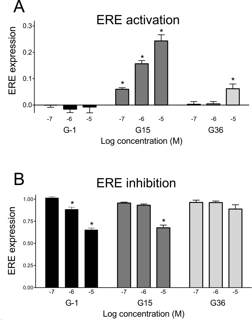
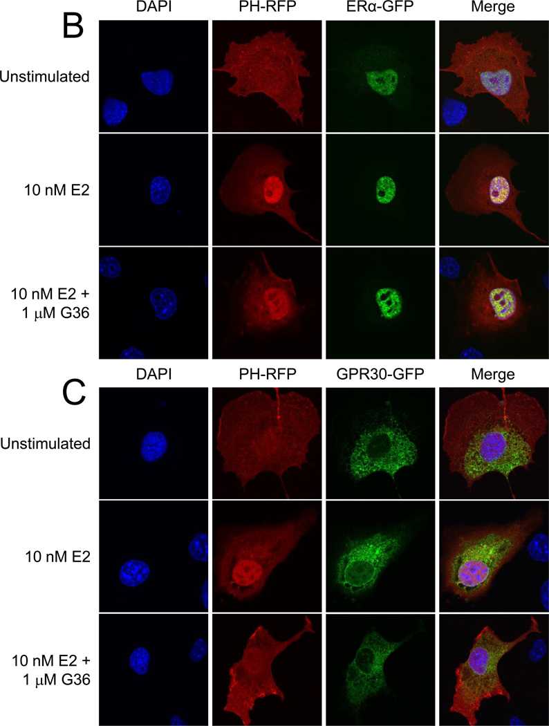
GPER/GPR30 is a seven-transmembrane G protein-coupled estrogen receptor that regulates many aspects of mammalian biology and physiology. We have previously described both a GPER-selective agonist G-1 and antagonist G15 based on a tetrahydro-3H-cyclopenta[c]quinoline scaffold. The antagonist lacks an ethanone moiety that likely forms important hydrogen bonds involved in receptor activation. Computational docking studies suggested that the lack of the ethanone substituent in G15 could minimize key steric conflicts, present in G-1, that limit binding within the ERα ligand binding pocket. In this report, we identify low-affinity cross-reactivity of the GPER antagonist G15 to the classical estrogen receptor ERα. To generate an antagonist with enhanced selectivity, we therefore synthesized an isosteric G-1 derivative, G36, containing an isopropyl moiety in place of the ethanone moiety. We demonstrate that G36 shows decreased binding and activation of ERα, while maintaining its antagonist profile towards GPER. G36 selectively inhibits estrogen-mediated activation of PI3K by GPER but not ERα. It also inhibits estrogen- and G-1-mediated calcium mobilization as well as ERK1/2 activation, with no effect on EGF-mediated ERK1/2 activation. Similar to G15, G36 inhibits estrogen- and G-1-stimulated proliferation of uterine epithelial cells in vivo. The identification of G36 as a GPER antagonist with improved ER counterselectivity represents a significant step towards the development of new highly selective therapeutics for cancer and other diseases.


Figures

Figure 1
Figure 1
Docking poses of selected ligands in the ER binding site: E2 green, G-1 magenta, G15 yellow. Arg394, which displays steric clashes with G-1 is depicted in orange in the lower left corner. The Pro399-Leu402 loop was hidden in order to better present the ligands.
Figure 2
Figure 2
High concentrations of G15 mediate weak activation of ERE. MCF-7 cells stably transfected with an ERE-GFP reporter were treated for 24 hours with the indicated concentration of E2, G-1 or G15. Whereas E2 shows half maximal activation at approximately 100 pM, G-1 shows no activation up to 10 µM. G15 exhibits limited activation (~15–20%) at concentrations ranging from 1–10 µM.
Figure 3
Figure 3
(A) Structures of estrogen (17β-estradiol) 1, G-1 2, G15 3 and G36 4. (B) Synthetic route for G36.
Figure 4
Figure 4
G36 exhibits improved binding selectivity towards ERα and ERβ compared to G15. Dose response profile of E2, G15 and G36 for competition of E2-Alexa633 binding to ERα-GFP (A) or ERβ-GFP (B). G36 shows decreased binding to ERα and ERβ at the highest concentration compared to G15.
Figure 5
Figure 5
G36 exhibits reduced activity towards ERE activation and inhibition compared to G15. (A) Activation of ERE-GFP response in MCF7 cells by increasing concentrations of G-1, G15 and G36. (B) Inhibition of ERE response induced by 1 nM E2 as a function of increasing concentrations of G-1, G15 and G36. *, p<0.05 vs. DMSO alone (A) or 1 nM E2 (B).
Figure 6
Figure 6
G36 inhibits E2-induced PI3K activation in cells expressing GPR30, but not in cells expressing ERα. (A) COS7 cells (which lack endogenous ERα, ERβ and GPER) transiently transfected with only PH-RFP show no activation of PI3K, as evidenced by the lack of nuclear translocation of PH-RFP, in response to E2, G-1 or G36 (all at 10 µM). (B) COS7 cells transiently transfected with ERα-GFP and PH-RFP activate PI3K, as evidenced by nuclear translocation of PH-RFP, in response to E2 and this response is not inhibited by the presence of G36. (C) In COS7 cells transiently transfected with GPR30-GFP and PH-RFP, G36 inhibits the PI3K activation induced by E2.
Figure 6
Figure 6
G36 inhibits E2-induced PI3K activation in cells expressing GPR30, but not in cells expressing ERα. (A) COS7 cells (which lack endogenous ERα, ERβ and GPER) transiently transfected with only PH-RFP show no activation of PI3K, as evidenced by the lack of nuclear translocation of PH-RFP, in response to E2, G-1 or G36 (all at 10 µM). (B) COS7 cells transiently transfected with ERα-GFP and PH-RFP activate PI3K, as evidenced by nuclear translocation of PH-RFP, in response to E2 and this response is not inhibited by the presence of G36. (C) In COS7 cells transiently transfected with GPR30-GFP and PH-RFP, G36 inhibits the PI3K activation induced by E2.
Figure 7
Figure 7
G36 inhibits E2 and G-1 mediated calcium mobilization in SKBr3 cells. (A) SKBr3 cells, which endogenously express only GPR30 but neither ERα nor ERβ, were monitored for calcium mobilization induced by either E2 (200 nM) or G-1 (200 nM). DMSO was added at the first arrow as a control and either E2 or G-1, as indicated at the second arrow. (B) Purinergic receptor activation (by 1 µM ATP, second arrow) was not inhibited by pretreatment with 10 µM G36 (first arrow). (C) E2- and G-1-mediated calcium mobilization are inhibited by increasing concentrations of G36 (pre-incubated for ~2 min, following the scheme in A). Data is normalized to E2 (200 nM) or G-1 (200 nM) activation of calcium mobilization in the presence of vehicle control (DMSO) as shown in A.
Figure 8
Figure 8
G36 inhibits ERK activation induced by E2 and G-1 via GPR30. SKBr3 cells were stimulated with E2 (10 nM), G-1 (10 nM) or EGF (10 ng/mL) in the presence of vehicle control (DMSO), 1 µM G15 or G36 (pre-incubated for 15 min) and analyzed for pERK and total ERK levels by Western blot. Quantitation of pERK activation was from three independent experiments. *, p<0.05 vs. DMSO alone.
Figure 9
Figure 9
G36 inhibits GPR30-mediated proliferation in vivo. Epithelial uterine cell proliferation was assessed in the presence of E2, G-1, G36, G-1 + G36 or E2 + G36. Amounts injected were as follows: E2: 10 ug/kg; G-1: 10 ug/kg; G36 (alone or in combination): 50 ug/kg. Ki-67 positivity of the uterine epithelium was determined by immunofluorescence microscopy. *, p<0.05 vs. sham; **, p<0.05 vs. E2.

References

    1. Edwards DP. Regulation of signal transduction pathways by estrogen and progesterone. Annu. Rev. Physiol. 2005;67:335–376. - PubMed
    1. Lange CA, Gioeli D, Hammes SR, Marker PC. Integration of rapid signaling events with steroid hormone receptor action in breast and prostate cancer. Annu. Rev. Physiol. 2007;69:171–199. - PubMed
    1. Prossnitz ER, Arterburn JB, Smith HO, Oprea TI, Sklar LA, Hathaway HJ. Estrogen signaling through the transmembrane G protein-coupled receptor GPR30. Annu. Rev. Physiol. 2008;70:165–190. - PubMed
    1. Albanito L, Madeo A, Lappano R, Vivacqua A, Rago V, Carpino A, Oprea TI, Prossnitz ER, Musti AM, Ando S, Maggiolini M. G protein-coupled receptor 30 (GPR30) mediates gene expression changes and growth response to 17beta-estradiol and selective GPR30 ligand G-1 in ovarian cancer cells. Cancer Res. 2007;67(4):1859–1866. - PubMed
    1. Pandey DP, Lappano R, Albanito L, Madeo A, Maggiolini M, Picard D. Estrogenic GPR30 signalling induces proliferation and migration of breast cancer cells through CTGF. EMBO J. 2009;28(5):523–532. - PMC - PubMed

Publication types

MeSH terms

LinkOut - more resources