Review
. 2012 Jan;1821(1):168-76.
doi: 10.1016/j.bbalip.2011.07.002. Epub 2011 Jul 12.

Signaling by vitamin A and retinol-binding protein in regulation of insulin responses and lipid homeostasis

Affiliations
Review

Signaling by vitamin A and retinol-binding protein in regulation of insulin responses and lipid homeostasis

Daniel C Berry et al. Biochim Biophys Acta. 2012 Jan.

Abstract

Vitamin A, retinol, circulates in blood bound to serum retinol binding protein (RBP) and is transported into cells by a membrane protein termed stimulated by retinoic acid 6 (STRA6). It was reported that serum levels of RBP are elevated in obese rodents and humans, and that increased level of RBP in blood causes insulin resistance. A molecular mechanism by which RBP can exert such an effect is suggested by the recent discovery that STRA6 is not only a vitamin A transporter but also functions as a surface signaling receptor. Binding of RBP-ROH to STRA6 induces the phosphorylation of a tyrosine residue in the receptor C-terminus, thereby activating a JAK/STAT signaling cascade. Consequently, in STRA6-expressing cells such as adipocytes, RBP-ROH induces the expression of STAT target genes, including SOCS3, which suppresses insulin signaling, and PPARγ, which enhances lipid accumulation. RBP-retinol thus joins the myriad of cytokines, growth factors and hormones which regulate gene transcription by activating cell surface receptors that signal through activation of Janus kinases and their associated transcription factors STATs. This article is part of a Special Issue entitled Retinoid and Lipid Metabolism.


Figures

Figure 1
Figure 1. Chemical structures of vitamin A (retinol), its precursors, and two important active metabolites
Retinol can be generated from β-carotene, present in plants, or from retinylesters, originating from animal sources. Retinol can then be metabolically transformed to active metabolites including all-trans-retinoic acid, which regulates gene transcription by activating the nuclear receptors RAR and PPARβ/δ, and 11-cis-retinal, which serves as a cofactor for the visual chromophore rhodopsin and is critical for vision.
Figure 2
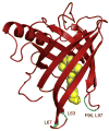
Figure 2. The three dimensional crystal structure of holo-retinol binding protein (RBP-ROH)
The human holo-RBP structure [23] (PDB ID 1BRP) was generated using Pymol (http://www.pymol.org/). The structure shows the eight stranded antiparallel β-sheet folded over itself to form a β-barrel. Retinol (yellow) is encapsulated by the barrel with the β-ionone ring buried in the binding pocket and the alcohol group is at the protein surface. Residues that stabilize the interactions of RBP with TTR are highlighted in blue. The location of these residues emphasize that interactions of RBP with TTR block the entrance to the RBP ligand-binding pocket.
Figure 3
Figure 3. The three dimensional crystal structure of the retinol-RBP-TTR complex
Human retinol-RBP-TTR [29] (PDB ID 1QAB) was generated using Pymol (http://www.pymol.org/). The TTR tetramer (magenta) is comprised of a dimer of dimers with two RBP molecules (red) bound to the opposite dimers. Interactions between RBP and TTR are mediated by residues at the entrance to the ligand binding pocket and span across the two TTR dimers.
Figure 4
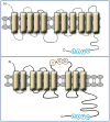
Figure 4. Alternative models for the structure of STRA6
Top: Computer model of STRA6 suggests that the protein is arranged in 11 transmembrane helices, a number of loops, and a large cytosolic domain. The phosphotyrosine motif in the cytosolic domain is highlighted. The model of STRA6 (GeneID 64220) was generated by the software http://bp.nuap.nagoya-u.ac.jp/sosui. Bottom: an alternative model in which STRA6 is arranged in 9 trans-membrane helices has been suggested [11]. It was proposed that highlighted residues in an extracellular loop located between helix 6 and 7 in this model stabilize the interactions of STRA6 with RBP [37].
Figure 5
Figure 5. Extracellular signalling molecules that utilize JAK/STAT pathways
The Table depicts known cytokines, hormones and growth factors that signal through cognate cell surface receptors to activate JAK/STAT signalling (adapted from Cell signaling, http://www.cellsignal.com/).
Figure 6
Figure 6. RBP-ROH activates STAT5 and induces STAT target genes to inhibit insulin signalling and enhance lipid accumulation
(a) Treatment of HepG2 cells with RBP-ROH triggers STAT5 phosphorylation. The effect is enhanced upon ectopic expression of STRA6 and abolished in the presence of a STRA6 lacking the SH2 binding motif. (b) RBP-ROH, but neither RBP nor ROH alone, induces the expression of the STAT target genes SOCS3 and PPARγ. (c) The ability of RBP-ROH to induce STAT target genes is not recapitulated by RBP-bound retinoic acid (RA) or retinal (RAL). (d) In cultured adipocytes, RBP-ROH suppresses the ability of insulin to trigger the phosphorylation of the insulin receptor, and does so in a STRA6-dependent fashion. (e) RBP-ROH enhances lipid accumulation in cultured adipocytes in a STRA6-depndent fashion. For details see [74].
Figure 7
Figure 7. Model of the RBP-ROH/STRA6/JAK/STAT pathway
Binding of RBP-ROH to the extracellular moiety of STRA6 triggers tyrosine phosphorylation in the receptor’s cytosolic domain. Phosphorylated STRA6 recruits and activates JAK2 which, in turn, phosphorylates STAT5. Activated STAT5 translocates to the nucleus to upregulate the expression of target genes, including SOCS3, which inhibits insulin signalling, and PPARγ, which enhances lipid accumulation.

Similar articles

Cited by

References

    1. McCollum EV, Davis M. The necessity of certain lipins in the diet during growth. J Biol Chem. 1913;15:167–175. - PubMed
    1. Osborne TB, Mendel LB. The vitamins in green foods. J Biol Chem. 1919;37:187–200.
    1. Noy N. Vitamin A. In: Stipanuk MH, editor. Biochemical, physiological, & molecular aspects of human nutrition. 2. Saunders Elsevier; St. Louis: 2006.
    1. Chambon P. A decade of molecular biology of retinoic acid receptors. Faseb J. 1996;10:940–954. - PubMed
    1. Germain P, Chambon P, Eichele G, Evans RM, Lazar MA, Leid M, De Lera AR, Lotan R, Mangelsdorf DJ, Gronemeyer H. International Union of Pharmacology. LX. Retinoic acid receptors. Pharmacol Rev. 2006;58:712–725. - PubMed

Publication types

MeSH terms