. 2011 Oct;21(5):686-93.
doi: 10.1016/j.sbi.2011.06.010. Epub 2011 Jul 21.

The power of correlative microscopy: multi-modal, multi-scale, multi-dimensional

Affiliations

The power of correlative microscopy: multi-modal, multi-scale, multi-dimensional

Jeffrey Caplan et al. Curr Opin Struct Biol. 2011 Oct.

Abstract

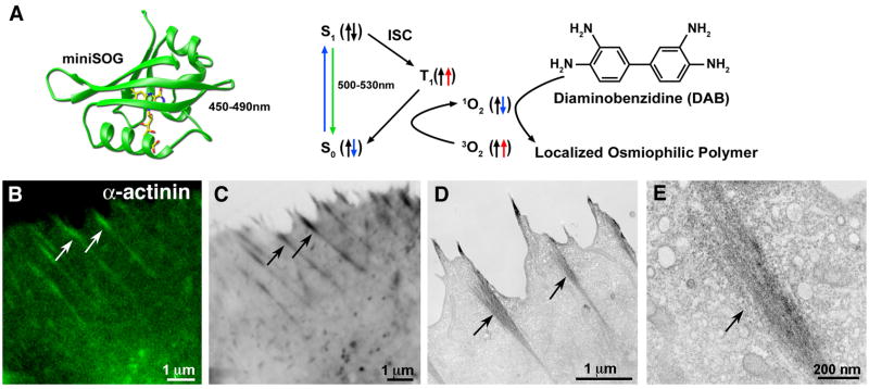
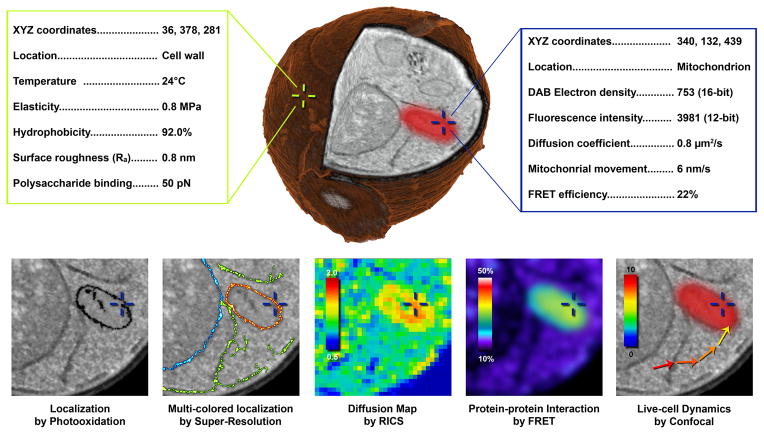
Correlative microscopy is a sophisticated approach that combines the capabilities of typically separate, but powerful microscopy platforms: often including, but not limited, to conventional light, confocal and super-resolution microscopy, atomic force microscopy, transmission and scanning electron microscopy, magnetic resonance imaging and micro/nano CT (computed tomography). When targeting rare or specific events within large populations or tissues, correlative microscopy is increasingly being recognized as the method of choice. Furthermore, this multi-modal assimilation of technologies provides complementary and often unique information, such as internal and external spatial, structural, biochemical and biophysical details from the same targeted sample. The development of a continuous stream of cutting-edge applications, probes, preparation methodologies, hardware and software developments will enable realization of the full potential of correlative microscopy.


Figures

Figure 1
Figure 1. Correlative light microscopy with TEM tomography
GFP-BIN1 expression in COS-1 cells. Bridging of specific GFP-BIN1 localization from confocal microscopy to high resolution TEM is illustrated by the correlation of a fluorescent image (A) with sequentially higher resolution EM micrographs (B–D). GFP-BIN1 localizes to tubules (arrow heads) and TEM places the localization into the subcellular context of mitochondria (M), lysosome (L) and nucleus (N) (D). An EM tomogram generated from 139 tilted images provides additional 3D information of the relation of tubules (yellow) to mitochondria, nuclear envelope, endosomal vesicles and lysosomes (grey), microtubules (red) and actin (black). Figure adapted from Speigelhalter et al. [8].
Figure 2
Figure 2. MiniSOG: a genetically encoded tag for correlative light and EM microscopy by photooxidation
(A) Structural model of miniSOG (left) and a diagram showing how blue-light excitation leads to photooxidation of diaminobenzidine (DAB) (right). (B–E) minSOG fused to α-actinin and expressed in HeLa cells. Confocal fluorescence (B) correlates with dark precipitation visible by transmitted light (C) and electron micrographs (D). Higher resolution EM shows discrete localization to a putative focal adhesion site (E). Arrows indicate correlated structures. Figure adapted from Shu and colleagues ([16].
Figure 3
Figure 3. Correlative AFM and fluorescence microcopy automated image alignment
Correlative microscopy using the Bruker BioScope Catalyst AFM/LM hybrid system with overlay alignment made in real time, pixel by pixel (A). User-defined locations can be targeted for single point force curves for measuring unbinding events (yellow crosses), AFM height image (brown ,35 × 35 μm overlay), or force volume mapping (yellow frame)and with the MIRO canvas fluorescence image can be directly chosen and navigated to using the software. Image provided courtesy of Alexandre Berquand (Bruker), Andreas Holloschi and Petra Kioschis (University of Applied Science). Marker (blue) previously found in an automatic finding stage (B). During registration, these AFM markers are mapped to the corresponding locations in the fluorescence image (C). The two images can then be automatically overlaid with high accuracy. The topography was generated from the AFM images and the colormap originates from the fluorescence image; the numerous bright “hills” indicate that the automatic alignment was successful (D). Panels B–D were provided courtesy of Serdar Cakici (University of North Carolina, Chapel Hill).
Figure 4
Figure 4. Hypothetical correlative approach to create an interactive and integrated 3D cellular map with biophysical, structural and chemical measurements acquired from a single yeast cell
Future prospects will likely include correlation of high resolution surface properties collected from different AFM modes (green text box) with characteristics obtained my multiple, advanced light microscopy or spectroscopy techniques (blue text box). This sample could then be rapidly frozen and processed to create high resolution 3D reconstructions from serial block-face imaging with focused ion beam SEM (center yeast cell). We envision this data set may be navigated in an interactive interface (similar to Google Earth), where specific points (cross hairs) can be chosen and a list of characteristics would be displayed (hypothetical values provided). The bottom row illustrates possibilities with such a muti-modal correlative dataset consisting of high resolution localization by DAB photooxidation, 3-color localization by STORM super resolution, diffusion coefficient map by raster image correlation spectroscopy, protein-protein interaction by FRET and live cell dynamics by confocal microscopy.

References

    1. Giepmans BN. Bridging fluorescence microscopy and electron microscopy. Histochemistry and cell biology. 2008;130:211–217. - PMC - PubMed
    1. Brown E, Verkade P. The use of markers for correlative light electron microscopy. Protoplasma. 2010;244:91–97. - PubMed
    1. Mironov AA, Beznoussenko GV. Correlative microscopy: a potent tool for the study of rare or unique cellular and tissue events. Journal of microscopy. 2009;235:308–321. - PubMed
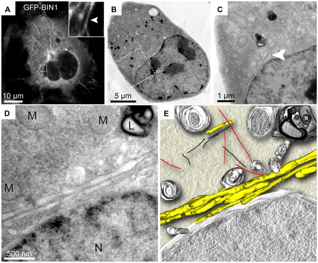
    1. Brown E, Mantell J, Carter D, Tilly G, Verkade P. Studying intracellular transport using high-pressure freezing and Correlative Light Electron Microscopy. Seminars in cell & developmental biology. 2009;20:910–919. With HeLa cells and combining live-cell time-lapse microscopy and high-pressure freezing to follow endocytosis, the authors showed merits of STEM over TEM for visualization of individual quantun dot and gold conjugated probes. - PubMed
    1. Muller-Reichert T, Mantler J, Srayko M, O'Toole E. Electron microscopy of the early Caenorhabditis elegans embryo. Journal of microscopy. 2008;230:297–307. - PubMed

Publication types

LinkOut - more resources