Review
. 2011 Nov;281(1-2):18-27.
doi: 10.1016/j.heares.2011.07.002. Epub 2011 Jul 18.

Apoptosis in acquired and genetic hearing impairment: the programmed death of the hair cell

Affiliations
Review

Apoptosis in acquired and genetic hearing impairment: the programmed death of the hair cell

Ken Op de Beeck et al. Hear Res. 2011 Nov.

Abstract

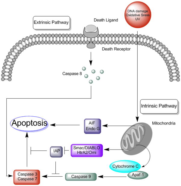
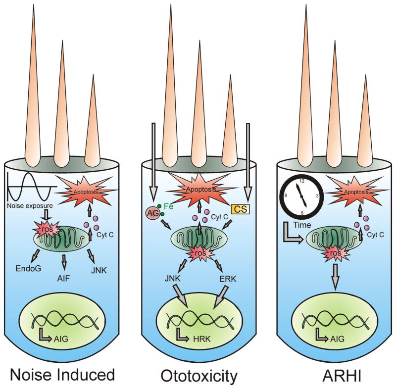
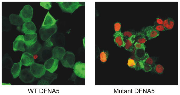
Apoptosis is an important physiological process. Normally, a healthy cell maintains a delicate balance between pro- and anti-apoptotic factors, allowing it to live and proliferate. It is thus not surprising that disturbance of this delicate balance may result in disease. It is a well known fact that apoptosis also contributes to several acquired forms of hearing impairment. Noise-induced hearing loss is the result of prolonged exposure to excessive noise, triggering apoptosis in terminally differentiated sensory hair cells. Moreover, hearing loss caused by the use of therapeutic drugs such as aminoglycoside antibiotics and cisplatin potentially may result in the activation of apoptosis in sensory hair cells leading to hearing loss due to the "ototoxicity" of the drugs. Finally, apoptosis is a key contributor to the development of presbycusis, age-related hearing loss. Recently, several mutations in apoptosis genes were identified as the cause of monogenic hearing impairment. These genes are TJP2, DFNA5 and MSRB3. This implies that apoptosis not only contributes to the pathology of acquired forms of hearing impairment, but also to genetic hearing impairment as well. We believe that these genes constitute a new functional class within the hearing loss field. Here, the contribution of apoptosis in the pathology of both acquired and genetic hearing impairment is reviewed.


Conflict of interest statement

Conflict of interest

The authors declare no conflict of interest.

Figures

Figure 1
Figure 1
Apoptosis can be induced through at least two pathways. The extrinsic pathway is activated through death receptors that reside on the plasma membrane. Binding of a death ligand to its receptor causes activation of caspase 8 that is then able to activate effector caspases such as caspase 3 and caspase 7. The intrinsic pathway on the other hand is activated from the inside of the cell. Stimuli such as DNA damage, oxidative stress and irradiation cause mitochondrial damage resulting in permeability changes of the outer mitochondrial membrane. This change causes the release of several pro apoptotic factors into the cytosol that trigger and amplify the apoptotic cascade. These proteins are then able to induce caspase-dependent and caspase-independent pathways. Release of endonuclease EndoG and apoptosis inducing factor (AIF) induce a caspase-independent apoptosis. On the other hand, release of cytochrome c proteins leads to oligomerization of apoptosis protease activating factor 1 (apaf-1) causing the formation of the so called apoptosome. This structure activates the initiator caspase 9, which in turn activates effector caspases such as caspase 3 and caspase 7. Meanwhile, Smac/DIABLO and HtrA2/Omi complexes counteract the inhibitory effects of inhibitor of apoptosis molecules (IAP) thereby enhancing activation of the apoptotic cascade.
Figure 2
Figure 2
Outer hair cells are vulnerable for different apoptosis-inducing stimuli. Depicted here are three outer hair cells that encounter various insults and as a result activate the apoptotic pathway. The left cell represents a noise-insulted cell. Noise exposure leads to an elevated generation of ROS, which causes mitochondria to release the pro-apoptotic factors EndoG and AIF. Moreover, ROS production is able to activate the JNK kinase system, leading to the transcription of several apoptosis inducing genes (AIG) in the nucleus. This causes the mitochondria to release cytochrome c and ultimately leads to apoptosis. In the case of ototoxicity, either aminoglycosides (AG) or cisplatin (CS) cause a rise in ROS formation. These ROS activate the JNK or ERK MAP kinase cascade, leading to transcription of AIG such as HRK (harakiri), which ultimately leads to activation of the apoptotic cell death program. The right cell depicts an ageing outer hair cell. The ageing theory predicts that in the course of time ROS concentration rises either due to depletion of antioxidant defenders or due to an elevated ROS formation. This causes mitochondrial damage and subsequent release of pro-apoptotic factors that finally induce apoptosis.
Figure 3
Figure 3
Confocal images of TUNEL stained HEK293T cells transfected with either WT or mutant DFNA5-EGFP. Red stained nuclei are TUNEL positive. Cells transfected with WT DFNA5 display a healthy appearance with little to no TUNEL positive nuclei. In contrast, cells transfected with mutant DFNA5 display an unhealthy appearance with clear TUNEL positive nuclei, indicative of apoptotic cell death.

References

    1. Ahmed ZM, Yousaf R, Lee BC, Khan SN, Lee S, Lee K, Husnain T, Rehman AU, Bonneux S, Ansar M, Ahmad W, Leal SM, Gladyshev VN, Belyantseva IA, Van Camp G, Riazuddin S, Friedman TB, Riazuddin S. Functional null mutations of MSRB3 encoding methionine sulfoxide reductase are associated with human deafness DFNB74. Am J Hum Genet. 2011;88:19–29. - PMC - PubMed
    1. Ahn JH, Kang HH, Kim YJ, Chung JW. Anti-apoptotic role of retinoic acid in the inner ear of noise-exposed mice. Biochem Biophys Res Commun. 2005;335:485–90. - PubMed
    1. Akino K, Toyota M, Suzuki H, Imai T, Maruyama R, Kusano M, Nishikawa N, Watanabe Y, Sasaki Y, Abe T, Yamamoto E, Tarasawa I, Sonoda T, Mori M, Imai K, Shinomura Y, Tokino T. Identification of DFNA5 as a target of epigenetic inactivation in gastric cancer. Cancer Sci. 2007;98:88–95. - PMC - PubMed
    1. Alam SA, Ikeda K, Oshima T, Suzuki M, Kawase T, Kikuchi T, Takasaka T. Cisplatin-induced apoptotic cell death in Mongolian gerbil cochlea. Hear Res. 2000;141:28–38. - PubMed
    1. Arslan E, Orzan E, Santarelli R. Global problem of drug-induced hearing loss. Ann N Y Acad Sci. 1999;884:1–14. - PubMed

Publication types