. 2011 Nov;44(2):223-30.
doi: 10.1016/j.nbd.2011.07.005. Epub 2011 Jul 18.

Morin attenuates tau hyperphosphorylation by inhibiting GSK3β

Affiliations

Morin attenuates tau hyperphosphorylation by inhibiting GSK3β

Eun Ji Gong et al. Neurobiol Dis. 2011 Nov.

Abstract

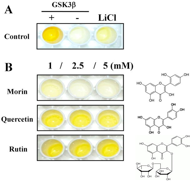
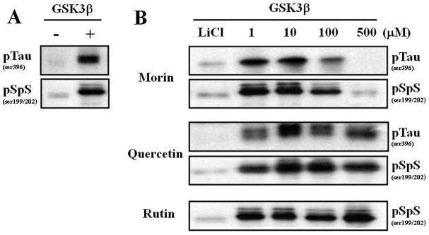
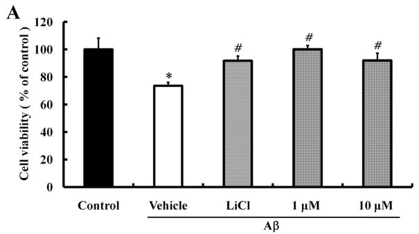
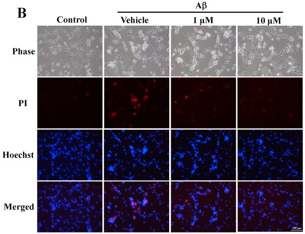
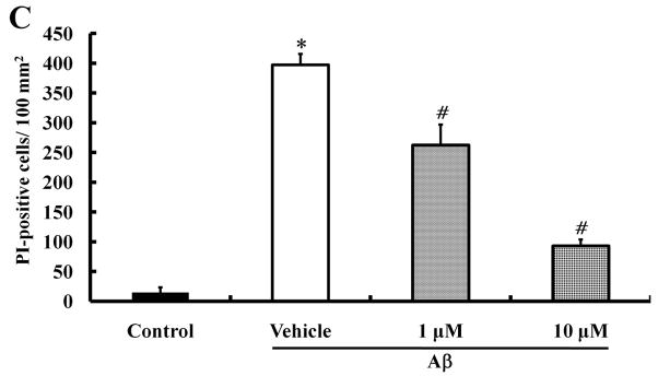
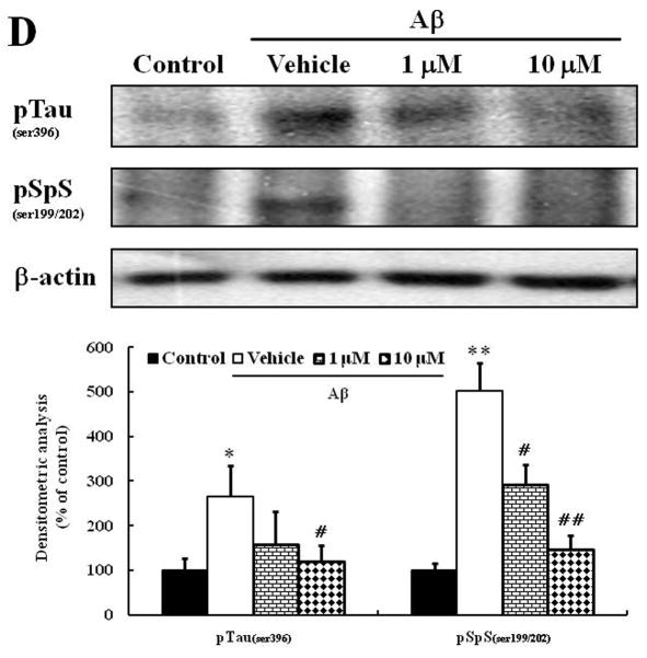
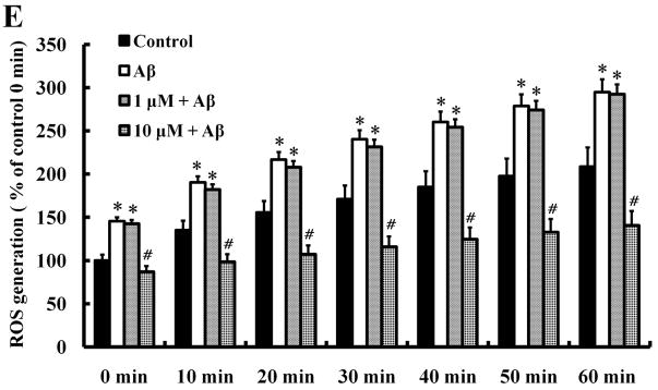
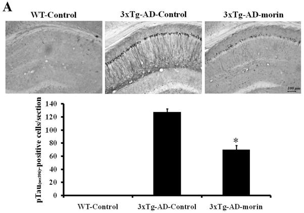
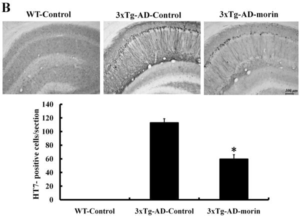
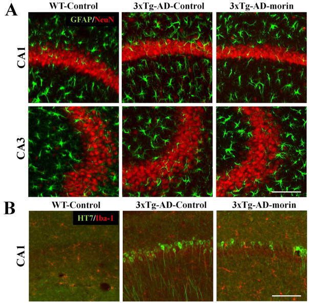
Alzheimer's disease (AD) is the major form of age-related dementia and is characterized by progressive cognitive impairment, the accumulation of extracellular amyloid β-peptide (Aβ), and intracellular hyperphosphorylated tau aggregates in affected brain regions. Tau hyperphosphorylation and accumulation in neurofibrillary tangles is strongly correlated with cognitive deficits, and is apparently a critical event in the dementia process because mutations in tau can cause a tangle-only form of dementia called frontotemporal lobe dementia. Among kinases that phosphorylate tau, glycogen synthase kinase 3β (GSK3β) is strongly implicated in AD pathogenesis. In the present study, we established an ELISA to screen for agents that inhibit GSK3β activity and found that the flavonoid morin effectively inhibited GSK3β activity and blocked GSK3β-induced tau phosphorylation in vitro. In addition, morin attenuated Aβ-induced tau phosphorylation and protected human neuroblastoma cells against Aβ cytotoxicity. Furthermore, treatment of 3xTg-AD mice with morin resulted in reductions in tau hyperphosphorylation and paired helical filament-like immunoreactivity in hippocampal neurons. Morin is a novel inhibitor of GSK3β that can reduce tau pathology in vivo and may have potential as a therapeutic agent in tauopathies.


Figures

Fig. 1
Fig. 1
Characterization of an ELISA to screen GSK3β inhibitors using β-catenin as a substrate. (A) Adding GSK3β significantly increased β-catenin phosphorylation, and LiCl (50 mM) effectively blocked GSK3β-induced phosphorylation. (B) Among flavonoids tested, we found that morin decreased the levels of phosphorylated β-catenin, while flavonoids with similar structures had no effect on GSK3β-induced phosphorylation.
Fig. 2
Fig. 2
Morin inhibits GSK3β activation-induced tau hyperphosphorylation in hippocampal homogenate. GSK3β-inhibitory effect of morin was evaluated by tau phosphorylation inhibition. Hippocampal tissue homogenate in 5X buffer with 1 mM ATP was pre-incubated with phytochemicals on ice for 30 minutes and then GSK3β was added and incubated at 37°C in a water bath for 30 minutes. Tau hyperphosphorylation (pTauser396, pSpSser199/202) was determined by immunoblot analysis. Note the molecular band shift due to increased phosphorylation. (A) GSK3β significantly increased tau hyperphosphorylation compared to homogenates incubated in the absence of GSK3β. (B) LiCl (50 mM), a well-known GSK3β inhibitor, decreased the tau hyperphosphorylation induced by GSK3β. Morin effectively decreased the GSK3β-mediated tau hyperphosphorylation in a concentration-dependent manner. In contrast, quercetin and rutin did not inhibit GSK3β-induced tau hyperphosphorylation.
Fig. 3
Fig. 3
Protective effects of morin against Aβ-induced neurotoxicity and tau hyperphosphorylation in human neuroblastoma cells. We investigated whether morin prevents Aβ-induced cell death and tau hyperphosphorylation in SH-SY5Y human neuroblastoma cells. (A) Cells were pretreated with morin (1 μM or 10 μM) and LiCl (100 μM) for 6 hours, followed by incubation with 10 μM Aβ for 24 hours. Cell viability was determined using the MTT assay. Pretreatment with morin and LiCl significantly increased cell viability in Aβ-treated cells. (B) We confirmed that morin protected against Aβ-induced cell death using Hoechst 33342 and PI staining. (C) Quantitative analysis of the number of PI-stained SH-SY5Y cells. The values are reported as the mean ± S.E.M (n=4). *p<0.01 compared to control. #p<0.01 compared to Aβ-treated culture. Scale bar = 100 μm. (D) Whole cell extracts from cells pretreated with morin for 6 hours, followed by treatment with 10 μM Aβ for 24 hours, were subjected to immunoblot analysis using antibodies against phosphorylated tau protein (pTauser396, pSpSser199/202). Level of β-actin was used as protein loading control. The blots were quantified by the densitometric analysis. (E) Total ROS were evaluated by DCF-DA method. Exposure to Aβ (10 μM) elevated ROS levels in the cells and morin treatment resulted in a significant attenuation of Aβ-induced ROS production. Values are the mean ± S.E.M (n=8). * p<0.01 compared to control. # p<0.01 compared to Aβ-treated culture (ANOVA with Fisher’s PLSD procedure).
Fig. 3
Fig. 3
Protective effects of morin against Aβ-induced neurotoxicity and tau hyperphosphorylation in human neuroblastoma cells. We investigated whether morin prevents Aβ-induced cell death and tau hyperphosphorylation in SH-SY5Y human neuroblastoma cells. (A) Cells were pretreated with morin (1 μM or 10 μM) and LiCl (100 μM) for 6 hours, followed by incubation with 10 μM Aβ for 24 hours. Cell viability was determined using the MTT assay. Pretreatment with morin and LiCl significantly increased cell viability in Aβ-treated cells. (B) We confirmed that morin protected against Aβ-induced cell death using Hoechst 33342 and PI staining. (C) Quantitative analysis of the number of PI-stained SH-SY5Y cells. The values are reported as the mean ± S.E.M (n=4). *p<0.01 compared to control. #p<0.01 compared to Aβ-treated culture. Scale bar = 100 μm. (D) Whole cell extracts from cells pretreated with morin for 6 hours, followed by treatment with 10 μM Aβ for 24 hours, were subjected to immunoblot analysis using antibodies against phosphorylated tau protein (pTauser396, pSpSser199/202). Level of β-actin was used as protein loading control. The blots were quantified by the densitometric analysis. (E) Total ROS were evaluated by DCF-DA method. Exposure to Aβ (10 μM) elevated ROS levels in the cells and morin treatment resulted in a significant attenuation of Aβ-induced ROS production. Values are the mean ± S.E.M (n=8). * p<0.01 compared to control. # p<0.01 compared to Aβ-treated culture (ANOVA with Fisher’s PLSD procedure).
Fig. 4
Fig. 4
Levels of tau hyperphosphorylation and HT-7 immunoreactivity were alleviated by morin treatment in the hippocampus of 3×Tg-AD mice. (A and B) Accumulation of tau phosphorylation and HT-7 in hippocampal neurons of vehicle- and morin-treated 3×Tg-AD mice were evaluated by immunohistochemistry using antibodies pTauser396 (A) and HT7 (B). Scale bar = 100 μm. Phospho-tau- and HT7-stained neurons were observed in CA1 region of 3×Tg-AD mice. However, morin treatment decreased the numbers of phospho-tau- and HT7-stained CA1 neurons. No staining was detected in WT mice. Value are the mean ± S.E.M (n=4). * p<0.01 compared to 3×Tg-AD-Control (ANOVA with Fisher’s PLSD procedure).
Fig. 4
Fig. 4
Levels of tau hyperphosphorylation and HT-7 immunoreactivity were alleviated by morin treatment in the hippocampus of 3×Tg-AD mice. (A and B) Accumulation of tau phosphorylation and HT-7 in hippocampal neurons of vehicle- and morin-treated 3×Tg-AD mice were evaluated by immunohistochemistry using antibodies pTauser396 (A) and HT7 (B). Scale bar = 100 μm. Phospho-tau- and HT7-stained neurons were observed in CA1 region of 3×Tg-AD mice. However, morin treatment decreased the numbers of phospho-tau- and HT7-stained CA1 neurons. No staining was detected in WT mice. Value are the mean ± S.E.M (n=4). * p<0.01 compared to 3×Tg-AD-Control (ANOVA with Fisher’s PLSD procedure).
Fig. 5
Fig. 5
Early tau hyperphosphorylation is not associated with neuronal loss or glial hyperreactivity in 3×Tg-AD mice. (A) To evaluate the neuronal loss/damage in 3×Tg-AD mice, double-label immunohistochemistry was performed with primary antibodies against a post-mitotic neuron marker NeuN (red) and the astrocyte marker GFAP (green). There was no sign of neuronal death and/or loss, or astroglial activation in the hippocampus. (B) Furthermore, microglia Iba-1 staining (red) showed no neuroinflammatory response in the hippocampus of 8 month-old 3×Tg-AD mice. However, we were able to confirm that morin significantly decreased HT7-stained CA1 neurons (green).

Similar articles

Cited by

References

    1. Alonso A, et al. Hyperphosphorylation induces self-assembly of tau into tangles of paired helical filaments/straight filaments. Proc Natl Acad Sci U S A. 2001;98:6923–8. - PMC - PubMed
    1. Alonso AC, et al. Alzheimer’s disease hyperphosphorylated tau sequesters normal tau into tangles of filaments and disassembles microtubules. Nat Med. 1996;2:783–7. - PubMed
    1. Alonso AC, et al. Role of abnormally phosphorylated tau in the breakdown of microtubules in Alzheimer disease. Proc Natl Acad Sci U S A. 1994;91:5562–6. - PMC - PubMed
    1. Alvarez G, et al. Regulation of tau phosphorylation and protection against beta-amyloid-induced neurodegeneration by lithium. Possible implications for Alzheimer’s disease. Bipolar Disord. 2002;4:153–65. - PubMed
    1. Arbel M, et al. Inhibition of amyloid precursor protein processing by beta-secretase through site-directed antibodies. Proc Natl Acad Sci U S A. 2005;102:7718–23. - PMC - PubMed

Publication types

MeSH terms